Journal article in Journal of Biomechanics:
Does energy consumption during walking increase with crouch severity among children with cerebral palsy?
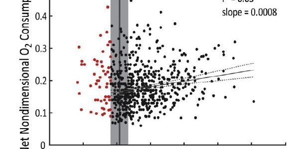
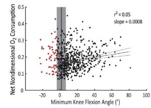 Abstract: Children with cerebral palsy (CP) expend more energy to walk compared to typically-developing peers. One of the most prevalent gait patterns among children with CP, crouch gait, is often singled out as especially exhausting. The dynamics of crouch gait increase external flexion moments and the demand on extensor muscles. This elevated demand is thought to dramatically increase energy expenditure. However, the impact of crouch severity on energy expenditure has not been investigated among children with CP. We evaluated oxygen consumption and gait kinematics for 573 children with bilateral CP. The average net nondimensional oxygen consumption during gait of the children with CP (0.18 ± 0.06) was 2.9 times that of speed-matched typically-developing peers. Crouch severity was only modestly related to oxygen consumption, with measures of knee flexion angle during gait explaining only 5–20% of the variability in oxygen consumption. While knee moment and muscle activity were moderately to strongly correlated with crouch severity (r2 = 0.13–0.73), these variables were only weakly correlated with oxygen consumption (r2 = 0.02–0.04). Thus, although the dynamics of crouch gait increased muscle demand, these effects did not directly result in elevated energy expenditure. In clinical gait analysis, assumptions about an individual’s energy expenditure should not be based upon kinematics or kinetics alone. Identifying patient-specific factors that contribute to increased energy expenditure may provide new pathways to improve gait for children with CP.

