KM Steele, MC Tresch, EJ Perreault, (2013) “The number and choice of muscles impact the results of muscle synergy analyses,” Frontiers in Computational Neuroscience

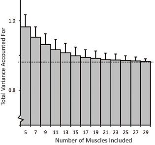
tVAF decreases with increasing number of muscles included in synergy analysis

Journal article accepted in Frontiers in Computational Neuroscience:

The number and choice of muscles impact the results of muscle synergy analyses

One theory for how humans control movement is that muscles are activated in weighted groups or synergies. Studies have shown that electromyography (EMG) from a variety of tasks can be described by a low-dimensional space thought to reflect synergies. These studies use algorithms, such as nonnegative matrix factorization, to identify synergies from EMG. Due to experimental constraints, EMG can rarely be taken from all muscles involved in a task. However, it is unclear if the choice of muscles included in the analysis impacts estimated synergies. The aim of our study was to evaluate the impact of the number and choice of muscles on synergy analyses. We used a musculoskeletal model to calculate muscle activations required to perform an isometric upper-extremity task. Synergies calculated from the activations from the musculoskeletal model were similar to a prior experimental study. To evaluate the impact of the number of muscles included in the analysis, we randomly selected subsets of between 5 and 29 muscles and compared the similarity of the synergies calculated from each subset to a master set of synergies calculated from all muscles. We determined that the structure of synergies is dependent upon the number and choice of muscles included in the analysis. When five muscles were included in the analysis, the similarity of the synergies to the master set was only 0.57 ± 0.54; however, the similarity improved to over 0.8 with more than ten muscles. We identified two methods, selecting dominant muscles from the master set or selecting muscles with the largest maximum isometric force, which significantly improved similarity to the master set and can help guide future experimental design. Analyses that included a small subset of muscles also over-estimated the variance accounted for (VAF) by the synergies compared to an analysis with all muscles. Thus, researchers should use caution using VAF to evaluate synergies when EMG is measured from a small subset of muscles. PDF

SR Hamner, A Seth, KM Steele, SL Delp, (2013) “A rolling constraint reduces ground reaction forces and moments in dynamic simulations of walking, running, and crouch gait.” Journal of Biomechanics

SR Hamner, A Seth, KM Steele, SL Delp, (2013) “A rolling constraint reduces ground reaction forces and moments in dynamic simulations of walking, running, and crouch gait.” Journal of Biomechanics

Journal article accepted in Journal of Biomechanics

A rolling constraint reduces ground reaction forces and moments in dynamic simulations of walking, running, and crouch gait

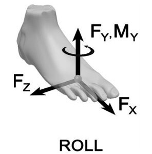
Recent advances in computational technology have dramatically increased the use of muscle-driven simulation to study accelerations produced by muscles during gait. Accelerations computed from muscle-driven simulations are sensitive to the model used to represent contact between the foot and ground. A foot-ground contact model must be able to calculate ground reaction forces and moments that are consistent with experimentally measured ground reaction forces and moments. We show here that a rolling constraint can model foot-ground contact and reproduce measured ground reaction forces and moments in an induced acceleration analysis of muscle-driven simulations of walking, running, and crouch gait. We also illustrate that a point constraint and a weld constraint used to model foot-ground contact in previous studies produce inaccurate reaction moments and lead to contradictory interpretations of muscle function. To enable others to use and test these different constraint types (i.e., rolling, point, and weld constraints) we have included them as part of an induced acceleration analysis in OpenSim, a freely-available biomechanics simulation package. PDF

KM Steele, A Seth, JL Hicks, MH Schwartz, SL Delp, (2013) “Muscle contributions to vertical and fore-aft accelerations are altered in subjects with crouch gait.” Gait & Posture

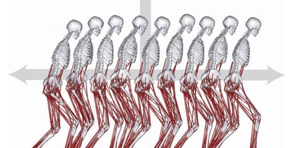
Musculoskeletal simulation used to evaluate crouch gait dynamics.

Journal article accepted in Gait & Posture:

Muscle contributions to vertical and fore-aft accelerations are altered in subjects with crouch gait.

The goals of this study were to determine if the muscle contributions to vertical and fore-aft acceleration of the mass center differ between crouch gait and unimpaired gait and if these muscle contributions change with crouch severity. Examining muscle contributions to mass center acceleration provides insight into the roles of individual muscles during gait and can provide guidance for treatment planning. We calculated vertical and fore-aft accelerations using musculoskeletal simulations of typically developing children and children with cerebral palsy and crouch gait. Analysis of these simulations revealed that during unimpaired gait the quadriceps produce large upward and backward accelerations during early stance, whereas the ankle plantarflexors produce large upward and forward accelerations later in stance. In contrast, during crouch gait, the quadriceps and ankle plantarflexors produce large, opposing fore-aft accelerations throughout stance. The quadriceps force required to accelerate the mass center upward was significantly larger in crouch gait than in unimpaired gait and increased with crouch severity. The gluteus medius accelerated the mass center upward during midstance in unimpaired gait; however, during crouch gait the upward acceleration produced by the gluteus medius was significantly reduced. During unimpaired gait the quadriceps and ankle plantarflexors accelerate the mass center at different times, efficiently modulating fore-aft accelerations. However, during crouch gait, the quadriceps and ankle plantarflexors produce fore-aft accelerations at the same time and the opposing fore-aft accelerations generated by these muscles contribute to the inefficiency of crouch gait. PDF

KM Steele, M van der Krogt, M Schwartz, SL Delp, (2012)“How much muscle strength is required to walk in a crouch gait?” Journal of Biomechanics

KM Steele, M van der Krogt, M Schwartz, SL Delp, (2012)“How much muscle strength is required to walk in a crouch gait?” Journal of Biomechanics

Journal article accepted in Journal of Biomechanics:

How much muscle strength is required to walk in a crouch gait?

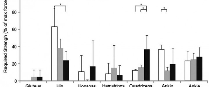
Muscle weakness is commonly cited as a cause of crouch gait in individuals with cerebral palsy; however, outcomes after strength training are variable and mechanisms by which muscle weakness may contribute to crouch gait are unclear. Understanding how much muscle strength is required to walk in a crouch gait compared to an unimpaired gait may provide insight into how muscle weakness contributes to crouch gait and assist in the design of strength training programs. The goal of this study was to examine how much muscle groups could be weakened before crouch gait becomes impossible. To investigate this question, we first created muscle-driven simulations of gait for three typically developing children and six children with cerebral palsy who walked with varying degrees of crouch severity. We then simulated muscle weakness by systematically reducing the maximum isometric force of each muscle group until the simulation could no longer reproduce each subject’s gait. This analysis indicated that moderate crouch gait required significantly more knee extensor strength than unimpaired gait. In contrast, moderate crouch gait required significantly less hip abductor strength than unimpaired gait, and mild crouch gait required significantly less ankle plantarflexor strength than unimpaired gait. The reduced strength required from the hip abductors and ankle plantarflexors during crouch gait suggests that weakness of these muscle groups may contribute to crouch gait and that these muscle groups are potential targets for strength training. PDF

KM Steele, M Demers, M Schwartz, SL Delp, (2012) “Compressive tibiofemoral forces during crouch gait.” Gait & Posture

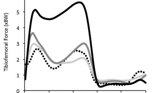
Tibiofemoral contact forces increases to over 5 times body-weight in severe crouch gait.

Journal article accepted in Gait & Posture:

Compressive tibiofemoral forces during crouch gait

Crouch gait, a common walking pattern in individuals with cerebral palsy, is characterized by excessive flexion of the hip and knee. Many subjects with crouch gait experience knee pain, perhaps because of elevated muscle forces and joint loading. The goal of this study was to examine how muscle forces and compressive tibiofemoral force change with the increasing knee flexion associated with crouch gait. Muscle forces and tibiofemoral force were estimated for three unimpaired children and nine children with cerebral palsy who walked with varying degrees of knee flexion. We scaled a generic musculoskeletal model to each subject and used the model to estimate muscle forces and compressive tibiofemoral forces during walking. Mild crouch gait (minimum knee flexion 20–35°) produced a peak compressive tibiofemoral force similar to unimpaired walking; however, severe crouch gait (minimum knee flexion > 50°) increased the peak force to greater than 6 times body-weight, more than double the load experienced during unimpaired gait. This increase in compressive tibiofemoral force was primarily due to increases in quadriceps force during crouch gait, which increased quadratically with average stance phase knee flexion (i.e., crouch severity). Increased quadriceps force contributes to larger tibiofemoral and patellofemoral loading which may contribute to knee pain in individuals with crouch gait. PDF