KM Steele, M van der Krogt, M Schwartz, SL Delp, (2012)“How much muscle strength is required to walk in a crouch gait?” Journal of Biomechanics

KM Steele, M van der Krogt, M Schwartz, SL Delp, (2012)“How much muscle strength is required to walk in a crouch gait?” Journal of Biomechanics

Journal article accepted in Journal of Biomechanics:

How much muscle strength is required to walk in a crouch gait?

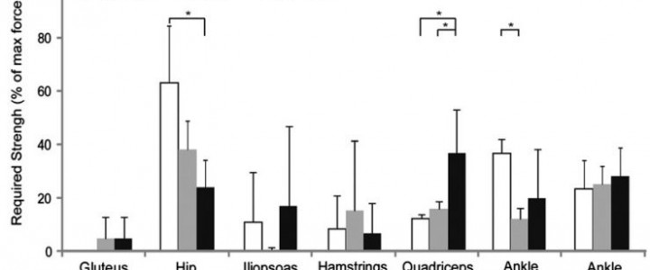
Muscle weakness is commonly cited as a cause of crouch gait in individuals with cerebral palsy; however, outcomes after strength training are variable and mechanisms by which muscle weakness may contribute to crouch gait are unclear. Understanding how much muscle strength is required to walk in a crouch gait compared to an unimpaired gait may provide insight into how muscle weakness contributes to crouch gait and assist in the design of strength training programs. The goal of this study was to examine how much muscle groups could be weakened before crouch gait becomes impossible. To investigate this question, we first created muscle-driven simulations of gait for three typically developing children and six children with cerebral palsy who walked with varying degrees of crouch severity. We then simulated muscle weakness by systematically reducing the maximum isometric force of each muscle group until the simulation could no longer reproduce each subject’s gait. This analysis indicated that moderate crouch gait required significantly more knee extensor strength than unimpaired gait. In contrast, moderate crouch gait required significantly less hip abductor strength than unimpaired gait, and mild crouch gait required significantly less ankle plantarflexor strength than unimpaired gait. The reduced strength required from the hip abductors and ankle plantarflexors during crouch gait suggests that weakness of these muscle groups may contribute to crouch gait and that these muscle groups are potential targets for strength training. PDF

KM Steele, M Demers, M Schwartz, SL Delp, (2012) “Compressive tibiofemoral forces during crouch gait.” Gait & Posture

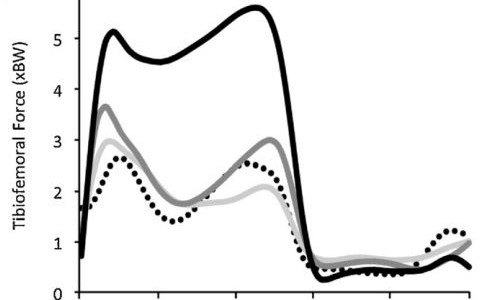
Tibiofemoral contact forces increases to over 5 times body-weight in severe crouch gait.

Journal article accepted in Gait & Posture:

Compressive tibiofemoral forces during crouch gait

Crouch gait, a common walking pattern in individuals with cerebral palsy, is characterized by excessive flexion of the hip and knee. Many subjects with crouch gait experience knee pain, perhaps because of elevated muscle forces and joint loading. The goal of this study was to examine how muscle forces and compressive tibiofemoral force change with the increasing knee flexion associated with crouch gait. Muscle forces and tibiofemoral force were estimated for three unimpaired children and nine children with cerebral palsy who walked with varying degrees of knee flexion. We scaled a generic musculoskeletal model to each subject and used the model to estimate muscle forces and compressive tibiofemoral forces during walking. Mild crouch gait (minimum knee flexion 20–35°) produced a peak compressive tibiofemoral force similar to unimpaired walking; however, severe crouch gait (minimum knee flexion > 50°) increased the peak force to greater than 6 times body-weight, more than double the load experienced during unimpaired gait. This increase in compressive tibiofemoral force was primarily due to increases in quadriceps force during crouch gait, which increased quadratically with average stance phase knee flexion (i.e., crouch severity). Increased quadriceps force contributes to larger tibiofemoral and patellofemoral loading which may contribute to knee pain in individuals with crouch gait. PDF

KM Steele, A Seth, JL Hicks, M Schwartz, SL Delp, (2010) “Muscle contributions to support during single-limb stance in crouch gait.” Journal of Biomechanics

KM Steele, A Seth, JL Hicks, M Schwartz, SL Delp,  (2010) “Muscle contributions to support during single-limb stance in crouch gait.” Journal of Biomechanics

Journal article accepted in Journal of Biomechanics:

Muscle contributions to support during single-limb stance in crouch gait

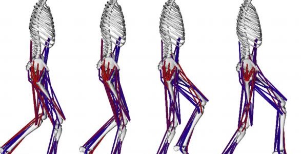
Pathological movement patterns like crouch gait are characterized by abnormal kinematics and muscle activations that alter how muscles support the body weight during walking. Individual muscles are often the target of interventions to improve crouch gait, yet the roles of individual muscles during crouch gait remain unknown. The goal of this study was to examine how muscles contribute to mass center accelerations and joint angular accelerations during single-limb stance in crouch gait, and compare these contributions to unimpaired gait. Subject-specific dynamic simulations were created for ten children who walked in a mild crouch gait and had no previous surgeries. The simulations were analyzed to determine the acceleration of the mass center and angular accelerations of the hip, knee, and ankle generated by individual muscles. The results of this analysis indicate that children walking in crouch gait have less passive skeletal support of body weight and utilize substantially higher muscle forces to walk than unimpaired individuals. Crouch gait relies on the same muscles as unimpaired gait to accelerate the mass center upward, including the soleus, vasti, gastrocnemius, gluteus medius, rectus femoris, and gluteus maximus. However, during crouch gait, these muscles are active throughout single-limb stance, in contrast to the modulation of muscle forces seen during single-limb stance in an unimpaired gait. Subjects walking in crouch gait rely more on proximal muscles, including the gluteus medius and hamstrings, to accelerate the mass center forward during single-limb stance than subjects with an unimpaired gait. PDF

DL Damiano, AS Arnold, KM Steele, S Delp, (2010) “Can strength training predictably improve gait kinematics in cerebral palsy?” Physical Therapy

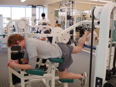
Hip and knee extension strength training exercises were used.

Journal article accepted in Physical Therapy:

Can strength training predictably improve gait kinematics in cerebral palsy?

Background: Computer simulations have demonstrated that excessive hip and knee flexion during gait, as frequently seen in ambulatory children with cerebral palsy (CP), can reduce the ability of muscles to provide antigravity support and increase the tendency of hip muscles to internally rotate the thigh. These findings suggest that therapies for improving upright posture during gait also may reduce excessive internal rotation. Objective: The goal of this study was to determine whether strength training can diminish the degree of crouched, internally rotated gait in children with spastic diplegic CP. Design: This was a pilot prospective clinical trial. Methods: Eight children with CP participated in an 8-week progressive resistance exercise program, with 3-dimensional gait analysis and isokinetic testing performed before and after the program. Secondary measures included passive range of motion, the Ashworth Scale, and the PedsQL CP Module. To identify factors that may have influenced outcome, individual and subgroup data were examined for patterns of change within and across variables. Results: Strength (force-generating capacity) increased significantly in the left hip extensors, with smaller, nonsignificant mean increases in the other 3 extensor muscle groups, yet kinematic and functional outcomes were inconsistent. The first reported subject-specific computer simulations of crouch gait were created for one child who showed substantial benefit to examine the factors that may have contributed to this outcome. Limitations: The sample was small, with wide variability in outcomes. Conclusions: Strength training may improve walking function and alignment in some patients for whom weakness is a major contributor to their gait deficits. However, in other patients, it may produce no change or even undesired outcomes. Given the variability of outcomes in this and other strengthening studies in CP, analytical approaches to determine the sources of variability are needed to better identify those individuals who are most likely to benefit from strengthening. PDF