Neuromechanics & Mobility Lab presents at NCM 2026

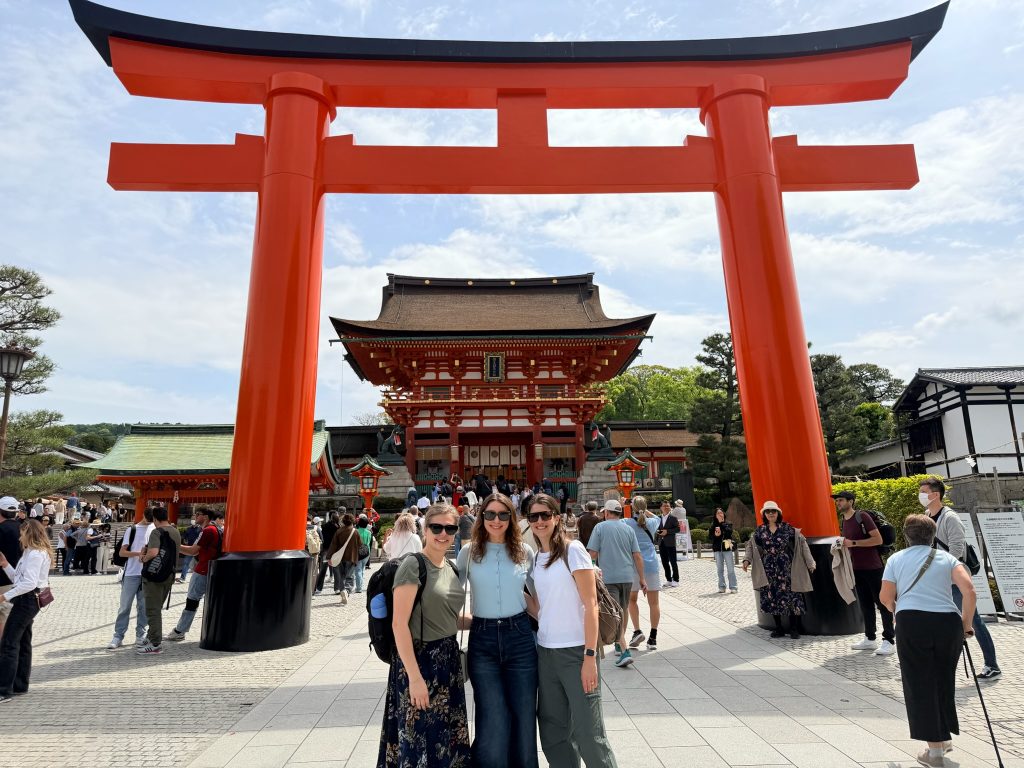
Members of the lab attended the Neural Control of Movement (NCM) conference in Kobe, Japan, bringing together an international community focused on understanding how the brain controls movement.

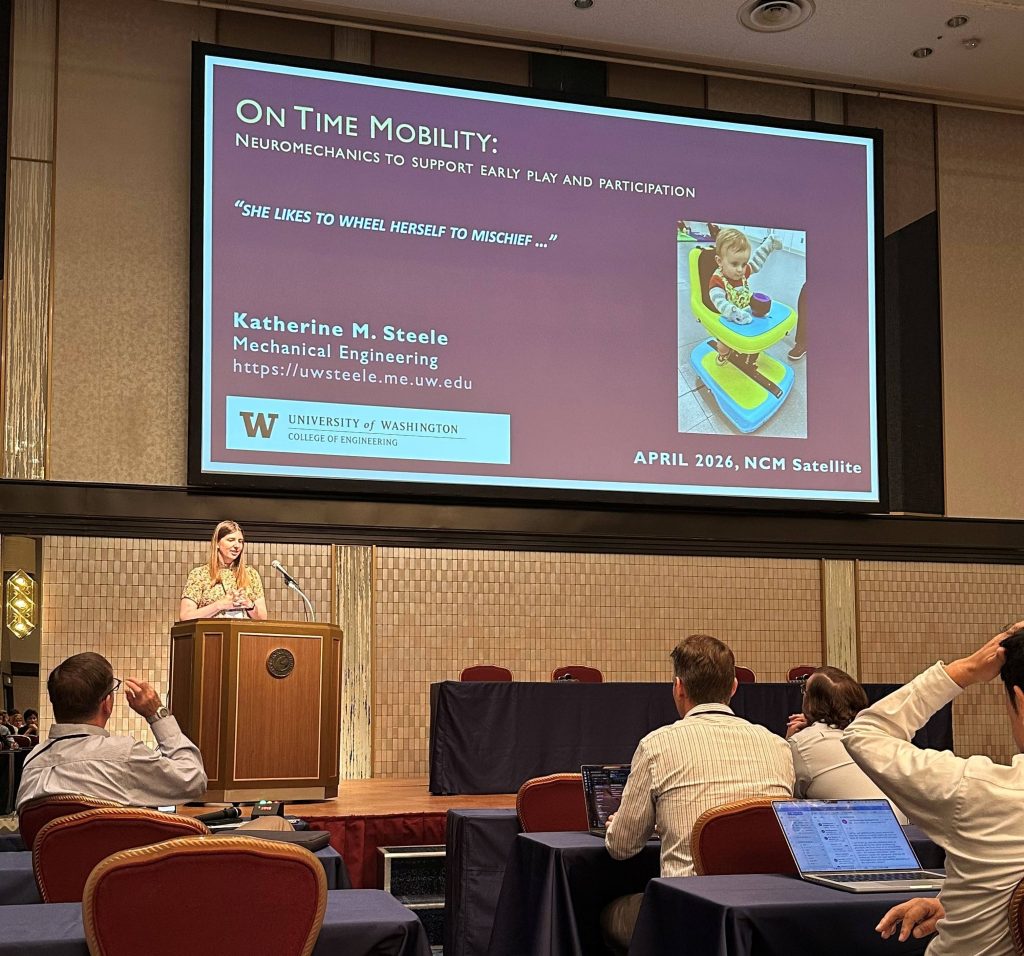
Dr. Kat Steele presented at the satellite meeting on “Precision neurorehabilitation for movement disorders: Integrating technology, neuroscience, and clinical practice,” where she also organized a session on gait and mobility. Her talk, “On time mobility: Neuromechanics to support early play and participation,” highlighted the lab’s work at the intersection of biomechanics and neurorehabilitation.

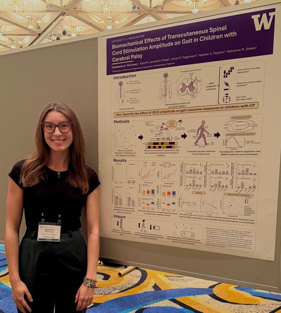
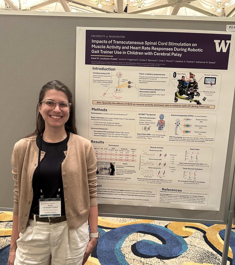
Madeleine McCreary presented a poster on “Biomechanical effects of transcutaneous spinal cord stimulation amplitude on gait in children with cerebral palsy.” And Katie Landwehr‑Prakel also presented a poster on “Impacts of transcutaneous spinal cord stimulation on muscle activity and heart rate responses during robotic gait trainer use in children with cerebral palsy.”

We were also excited to connect with lab alum and collaborator Dr. Charlotte DeVol, highlighting the strong ongoing collaborations within our broader research community.

It was an exciting opportunity to share research, connect with collaborators, and engage with the global NCM community.

  • Three people pose in front of a large red torii gate at a busy shrine in Japan, with a traditional temple building and visitors in the background.
  • Dr. Kat Steele stands at a podium presenting to an audience, with a large slide reading “On Time Mobility: Neuromechanics to support early play and participation” displayed on a screen at an NCM conference session.
  • Katie stands in front of a scientific poster titled “Impacts of Transcutaneous Spinal Cord Stimulation on Muscle Activity and Heart Rate Responses During Robotic Gait Trainer Use in Children with Cerebral Palsy” at a poster session.

HA Feldner, C Papazian, KM Peters, CJ Cruetzfeldt, KM Steele (2021) “Clinical Use of Surface Electromyography to Track Acute Upper Extremity Muscle Recovery after Stroke: A Descriptive Case Study of a Single Patient”

Journal Article in Applied System Innovation:

This work highlights the potential of wearable technologoies to monitor muscle activity changes during stroke recovery in acute clinical settings and their importance for motivation and understanding of progression from the survivor’s point of view: ‘I was hopeful that it would show signs of things that are occurring when I couldn’t physically feel it…if you had other scientific evidence that things were happening, even beyond their notion that it would, it gives you a lot of hope. You just have to be patient, and it’s harder to take when someone tells you, but easier to understand if someone actually shows you’.

Left image depicts arm with pads placed over muscle with right pictures depicting similar image

Aim: Describe the use of wireless sEMG sensors to examine changes in muscle activity during acute and subacute phases of stroke recovery, and understand the participant’s perceptions of sEMG monitoring.

Method: Muscle activity was tracked by five wireless sEMG sensors beginning three days post-stroke and continued through discharge from inpatient rehabilitation. Activity logs were completed each session, and a semi-structured interview occurred at the final session with three- and eight-month follow-up sessions.

Results: The longitudinal monitoring of muscle and movement recovery in the clinic and community was feasible using sEMG sensors. The participant and medical team felt monitoring was unobtrusive, interesting, and motivating for recovery, but desired greater in-session feedback to inform rehabilitation.

Interpretation: This work highlights that barriers in equipment and signal quality still exist, but capitalizing on wearable sensing technology in the clinic holds promise for enabling personalized stroke recovery.