KM Steele, MC Tresch, EJ Perreault (2015) “Consequences of biomechanically constrained tasks in the design and interpretation of synergy analyses.” Journal of Neurophysiology

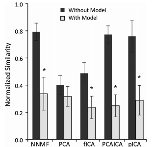
Synergy similarity is reduced with musculoskeletal constraints.

Journal article in Journal of Neurophysiology

Consequences of biomechanically constrained tasks in the design and interpretation of synergy analyses

Matrix factorization algorithms are commonly used to analyze muscle activity and provide insight into neuromuscular control. These algorithms identify low-dimensional subspaces, commonly referred to as synergies, which can describe variation in muscle activity during a task. Synergies are often interpreted as reflecting underlying neural control; however, it is unclear how these analyses are influenced by biomechanical and task constraints, which can also lead to low-dimensional patterns of muscle activation. The aim of this study was to evaluate whether commonly used algorithms and experimental methods can accurately identify synergy-based control strategies. This was accomplished by evaluating synergies from five common matrix factorization algorithms using muscle activations calculated from 1) a biomechanically constrained task using a musculoskeletal model and 2) without task constraints using random synergy activations. Algorithm performance was assessed by calculating the similarity between estimated synergies and those imposed during the simulations; similarities ranged from 0 (random chance) to 1 (perfect similarity). Although some of the algorithms could accurately estimate specified synergies without biomechanical or task constraints (similarity >0.7), with these constraints the similarity of estimated synergies decreased significantly (0.3-0.4). The ability of these algorithms to accurately identify synergies was negatively impacted by correlation of synergy activations, which are increased when substantial biomechanical or task constraints are present. Increased variability in synergy activations, which can be captured using robust experimental paradigms that include natural variability in motor activation patterns, improved identification accuracy but did not completely overcome effects of biomechanical and task constraints. These results demonstrate that a biomechanically constrained task can reduce the accuracy of estimated synergies and highlight the importance of using experimental protocols with physiological variability to improve synergy analyses. PDF

KM Steele and S Lee (2014) “Using dynamic musculoskeletal simulation to evaluate altered muscle properties in cerebral palsy.” Proceedings of ASME Dynamics Systems and Control

KM Steele and S Lee (2014) “Using dynamic musculoskeletal simulation to evaluate altered muscle properties in cerebral palsy.” Proceedings of ASME Dynamics Systems and Control

Paper accepted at ASME Dynamics Systems and Control Conference:

Using dynamic musculoskeletal simulation to evaluate altered muscle properties in cerebral palsy

Abstract: Cerebral palsy is caused by an injury to the brain, but also causes many secondary changes in the musculoskeletal system. Altered muscle properties such as contracture, an increased passive resistance to stretch, are common but vary widely between individuals and between muscles. Quantifying these changes is important to understand pathologic movement and create patient-specific treatment plans. Musculoskeletal modeling and simulation have increasingly been used to evaluate pathologic movement in CP; however, these models are based upon muscle properties of unimpaired individuals. In this study, we used a dynamic musculoskeletal simulation of a simple motion, passively moving the ankle, to determine (1) if a model based upon unimpaired muscle properties can accurately represent individuals with cerebral palsy, and (2) if an optimization can be used to adjust passive muscle properties and characterize magnitude of contracture in individual muscles. We created musculoskeletal simulations of ankle motion for nine children with cerebral palsy. Results indicate that the unimpaired musculoskeletal model cannot accurately characterize passive ankle motion for most subjects, but adjusting tendon slack lengths can reduce error and help identify the magnitude of contracture for different muscles.

KM Steele, D Gaebler-Spira, S Lee, “Quantifying altered muscle properties with ultrasound elastography and musculoskeletal simulation.” Gait & Clinical Movement Analysis Society (Newark, Delaware) June 24-27, 2014.

KM Steele, D Gaebler-Spira, S Lee, “Quantifying altered muscle properties with ultrasound elastography and musculoskeletal simulation.” Gait & Clinical Movement Analysis Society (Newark, Delaware) June 24-27, 2014.

Kat Steele presents at Gait & Clinical Movement Analysis Society Conference:

Quantifying altered muscle properties with ultrasound elastography and musculoskeletal simulation

Newark, DE (June 24-27, 2014)


R Klemetti, KM Steele, P Moilanen, J Avela, J Timonen, (2014) “Contributions of individual muscles to the sagittal- and frontal-plane angular accelerations of the trunk in walking.” Journal of Biomechanics

R Klemetti, KM Steele, P Moilanen, J Avela, J Timonen, (2014) “Contributions of individual muscles to the sagittal- and frontal-plane angular accelerations of the trunk in walking.” Journal of Biomechanics

Journal article accepted in Journal of Biomechanics:

Contributions of individual muscles to the sagittal- and frontal-plane angular accelerations of the trunk in walking.

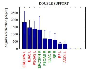
This study was conducted to analyze the unimpaired control of the trunk during walking. Studying the unimpaired control of the trunk reveals characteristics of good control. These characteristics can be pursued in the rehabilitation of impaired control. Impaired control of the trunk during walking is associated with aging and many movement disorders. This is a concern as it is considered to increase fall risk. Muscles that contribute to the trunk control in normal walking may also contribute to it under perturbation circumstances, attempting to prevent an impending fall. Knowledge of such muscles can be used to rehabilitate impaired control of the trunk. Here, angular accelerations of the trunk induced by individual muscles, in the sagittal and frontal planes, were calculated using 3D muscle-driven simulations of seven young healthy subjects walking at free speed. Analysis of the simulations demonstrated that the abdominal and back muscles displayed large contributions throughout the gait cycle both in the sagittal and frontal planes. Proximal lower-limb muscles contributed more than distal muscles in the sagittal plane, while both proximal and distal muscles showed large contributions in the frontal plane. Along with the stance-limb muscles, the swing-limb muscles also exhibited considerable contribution. The gluteus medius was found to be an important individual frontal-plane control muscle; enhancing its function in pathologies could ameliorate gait by attenuating trunk sway. In addition, since gravity appreciably accelerated the trunk in the frontal plane, it may engender excessive trunk sway in pathologies. PDF