Journal article in Clinical Biomechanics:
Kat Steele partnered with Sabrina Lee from Northwestern University and the Rehabilitation Institute of Chicago to investigate shearwave ultrasound elastography as a new tool to quantify changes in muscle properties in cerebral palsy.
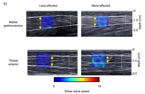 Abstract: Individuals with cerebral palsy tend to have altered muscle architecture and composition, but little is known about the muscle material properties, specifically stiffness. Shear wave ultrasound elastography allows shear wave speed, which is related to stiffness, to be measured in vivo in individual muscles. Our aim was to evaluate the material properties, specifically stiffness, as measured by shear wave speed of the medial gastrocnemius and tibialis anterior muscles in children with hemiplegic cerebral palsy across a range of ankle torques and positions, and fascicle strains. Shear wave speed was measured bilaterally in the medial gastrocnemius and tibialis anterior over a range of ankle positions and torques using shear wave ultrasound elastography in eight individuals with hemiplegic cerebral palsy. B-mode ultrasound was used to measure muscle thickness and fascicle strain. Shear waves traveled faster in the medial gastrocnemius and tibialis anterior of the more-affected limb by 14% (P = 0.024) and 20% (P = 0.03), respectively, when the ankle was at 90°. Shear wave speed in the medial gastrocnemius increased as the ankle moved from plantarflexion to dorsiflexion (less affected: r2 = 0.82, P < 0.001; more-affected: r2 = 0.69, P < 0.001) and as ankle torque increased (less affected: r2 = 0.56,P < 0.001; more-affected: r2 = 0.45, P < 0.001). In addition, shear wave speed was strongly correlated with fascicle strain (less affected: r2 = 0.63, P < 0.001; more-affected: r2 = 0.53, P < 0.001). The higher shear wave speed in the more-affected limb of individuals with cerebral palsy indicates greater muscle stiffness, and demonstrates the clinical potential of shear wave elastography as a non-invasive tool for investigating mechanisms of altered muscle properties and informing diagnosis and treatment.