M Yamagami, KM Peters, I Milovanovic, I Kuang, Z Yang, N Lu, KM Steele (2018) “Assessment of Dry Epidermal Electrodes for Long-Term Electromyography Measurements.” Sensors

Sample sEMG signal from one subject’s FCU for (left) MVIC; (middle) dynamic and (right) functional tests indicate that there were no significant differences between the Delsys (lighter grey) and ESS electrodes (darker grey) based on raw sEMG amplitude, linear envelope amplitude, or power spectral density.

Journal article in Sensors:

In collaboration with University of Texas – Austin, we evaluated a new flexible, gold-based epidermal electrode for sensing muscle activity.

Sample sEMG signal from one subject’s FCU for (left) MVIC; (middle) dynamic and (right) functional tests indicate that there were no significant differences between the Delsys (lighter grey) and ESS electrodes (darker grey) based on raw sEMG amplitude, linear envelope amplitude, or power spectral density.Background: Commercially available electrodes can only provide quality surface electromyography (sEMG) measurements for a limited duration due to user discomfort and signal degradation, but in many applications, collecting sEMG data for a full day or longer is desirable to enhance clinical care. Few studies for long-term sEMG have assessed signal quality of electrodes using clinically relevant tests. The goal of this research was to evaluate flexible, gold-based epidermal sensor system (ESS) electrodes for long-term sEMG recordings.

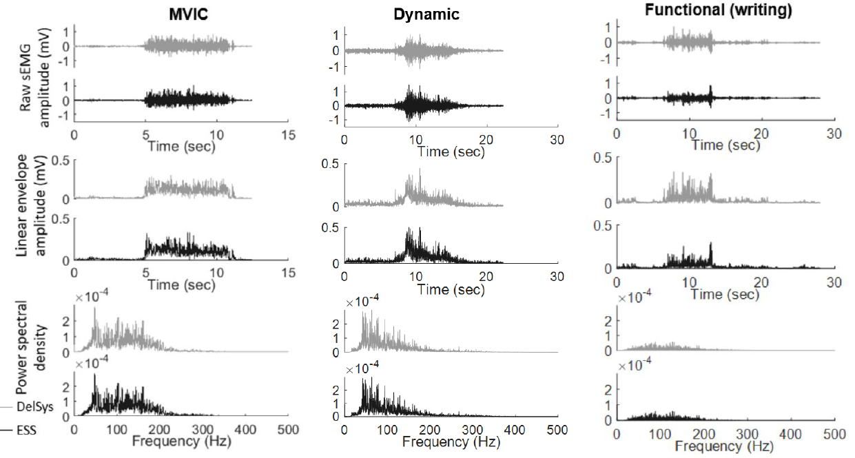
Methods: We collected sEMG and impedance data from eight subjects from ESS and standard clinical electrodes on upper extremity muscles during maximum voluntary isometric contraction tests, dynamic range of motion tests, the Jebsen Taylor Hand Function Test, and the Box & Block Test. Four additional subjects were recruited to test the stability of ESS signals over four days.

Results: Signals from the ESS and traditional electrodes were strongly correlated across tasks. Measures of signal quality, such as signal-to-noise ratio and signal-to-motion ratio, were also similar for both electrodes.

Conclusions: Over the four-day trial, no significant decrease in signal quality was observed in the ESS electrodes, suggesting that thin, flexible electrodes may provide a robust tool that does not inhibit movement or irritate the skin for long-term measurements of muscle activity in rehabilitation and other applications.

KM Steele, BR Shuman, MH Schwartz (2017) 
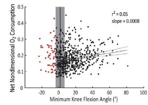
“Crouch severity is a poor predictor of elevated oxygen consumption in cerebral palsy.” Journal of Biomechanics

Scatter plot illustrating that there is not a significant correlation between minimum knee flexion angle during stance and oxygen consumption.

Journal article in Journal of Biomechanics:

Does energy consumption during walking increase with crouch severity among children with cerebral palsy?

Scatter plot illustrating that there is not a significant correlation between minimum knee flexion angle during stance and oxygen consumption.Abstract: Children with cerebral palsy (CP) expend more energy to walk compared to typically-developing peers. One of the most prevalent gait patterns among children with CP, crouch gait, is often singled out as especially exhausting. The dynamics of crouch gait increase external flexion moments and the demand on extensor muscles. This elevated demand is thought to dramatically increase energy expenditure. However, the impact of crouch severity on energy expenditure has not been investigated among children with CP. We evaluated oxygen consumption and gait kinematics for 573 children with bilateral CP. The average net nondimensional oxygen consumption during gait of the children with CP (0.18 ± 0.06) was 2.9 times that of speed-matched typically-developing peers. Crouch severity was only modestly related to oxygen consumption, with measures of knee flexion angle during gait explaining only 5–20% of the variability in oxygen consumption. While knee moment and muscle activity were moderately to strongly correlated with crouch severity (r2 = 0.13–0.73), these variables were only weakly correlated with oxygen consumption (r2 = 0.02–0.04). Thus, although the dynamics of crouch gait increased muscle demand, these effects did not directly result in elevated energy expenditure. In clinical gait analysis, assumptions about an individual’s energy expenditure should not be based upon kinematics or kinetics alone. Identifying patient-specific factors that contribute to increased energy expenditure may provide new pathways to improve gait for children with CP.

NIH cerebral palsy strategic plan – our comments

The National Institutes of Health recently released the “Strategic Plan for Cerebral Palsy Research” which outlines challenges and priorities to guide future research to improve the lives of people with cerebral palsy.

Our diverse research group enjoyed reading and discussing this plan, which will likely influence our future research goals and support. We’ve shared our group’s comments, organized and prepared by Dr. Heather Feldner, below:

“Our research group appreciated the committee’s focus on creating a centralized data source for CP, attention to the needs and perspectives of adults with CP, their childhood experiences, and their transition from pediatric to adult healthcare providers, and the call for greater caregiver support services and patient-reported outcomes. However, we also had concerns. First, the terminology is inconsistent and often inappropriate. “Cure”, “damage”, and the implication that people with CP cannot be “healthy” is not empowering language in supporting the lives, unique contributions, and perspectives of people with CP as diverse and valued individuals in our society. Further, while advocates of people with CP were included in this stakeholder group, there is a concerning lack of people who actually have a diagnosis of CP, when these should be the primary stakeholders setting a research agenda about their own lives and needs. Finally, given the uncertainty of government funding agencies like the NIH under the current administration’s budget proposal, and the speed of science of translating research from bench to bedside, it appears that too little priority has been placed on interventions or programs that could have an influence right now for the people living with CP in the US dealing with self-identified participation issues such as access to employment and education, as well as impairment-related needs such as pain management, access to technology, and functional mobility.

We are excited that NIH is engaged to set a national research agenda for cerebral palsy and we look forward to continuing to serve this community.

Logo of NINDS/NICHD Plan for cerebral palsy research

B Soran, L Lowes, KM Steele (2016) “Evaluation of infants with spinal muscular atrophy using convolutional neural networks.” European Conference on Computer Vision

Experimental set-up with infant positioned below Kinect depth camera.

Peer-reviewed paper at European Conference on Computer Vision:

30-second videos from a depth camera can be used in the evaluation of infants with spinal muscular atrophy.

Experimental set-up with infant positioned below Kinect depth camera.Abstract: Spinal Muscular Atrophy is the most common genetic cause of infant death. Due to its severity, there is a need for methods for automated estimation of disease progression. In this paper we propose a Convolutional-Neural-Network (CNN) model to estimate disease progression during infants’ natural behavior. With the proposed methodology, we were able to predict each child’s score on current behavior-based clinical exams with an average per-subject error of 6.96 out of 72 points (<10 % difference), using 30-second videos in leave-one-subject-out-cross-validation setting. When simple statistics were used over 30-second video-segments to estimate a score for longer videos, we obtained an average error of 5.95 (8 % error rate). By showing promising results on a small dataset (N = 70, 2-minute samples, which were handled as 1487, 30-second video segments), our methodology demonstrates that it is possible to benefit from CNNs on small datasets by proper design and data handling choices.

Can Technology Make a Difference in Pediatric Rehabilitation? – A NCMRR Webcast

Interested in how technology can be used to make a difference in pediatric rehabilitation? A video cast from the National Center for Medical Rehabilitation Research (NCMRR) discusses the topic in Bethesda MD. The workshop is organized by the Motion Analysis Laboratory and supported by the National Science Foundation and the National Institutes of Health.

The workshop on August 9th, 2016 brought together a group of experts in rehabilitation to discuss how technology can help us to address pressing needs in pediatric rehabilitation. To follow all of the talks this past week and listen to “Can Technology Make a Difference in Pediatric Rehabilitation?”, follow this link, CLICK HERE.