B Nguyen, N Baicoianu, D Howell, KM Peters, KM Steele (2020) “Accuracy and repeatability of smartphone sensors for measuring shank-to-vertical angle” Prosthetics & Orthotics International

Journal Article in Prosthetics & Orthotics International

Example of how the smartphone app was used for this research. The top images show a black smartphone attached with a running arm band to the side or front of the shank - the two positions tested in this research. The middle figure shows the placement of the reflective markers for 3D motion analysis to evaluate the accuracy of the smartphone measurements. Markers were placed on the lateral epicondyle of the knee, lateral maleolus of the ankle, tibial tuberosity, and the distal tibia. Blacklight was used to mark the position of each marker and hide the position from the clinicians. The bottom panel shows screenshots from the app. The first screen is used to align the device and has arrows at the top and bottom that remind the clinician which anatomical landmarks should be used to align the device while displaying the shank-to-vertical angle in real time. The second screenshot shows an example of the calculated shank-to-vertical angle while someone was walking. The average is shown with a bold black line, with all other trials shown in blue and excluded trials (e.g., when someone was stopping or turning) that deviated more than one standard deviation from other trials are shown in red. There is also text below the graph that provides summary measures, like shank-to-vertical angle in mid stand and cadence (steps/min). The results can be exported as a picture or sent via e-mail using the app.
A) Smartphone positioning on the front or side of the shank. B) Reflective markers on the the tibial tuberosity (TT) – distal tibia (DT) and lateral epicondyle (LE) – lateral malleolus (LM) were used to compare the accuracy of the smartphone to traditional motion capture. UV markings were used to keep placement of these markers constant while blinding clinicians. C) Sample screenshots of the mobile application, including the set-up screen and results automatically produced after a walking trial.

Background

Assessments of human movement are clinically important. However, accurate measurements are often unavailable due to the need for expensive equipment or intensive processing. For orthotists and therapists, shank-to-vertical angle (SVA) is one critical measure used to assess gait and guide prescriptions. Smartphone-based sensors may provide a widely-available platform to expand access to quantitative assessments.

Objectives

Assess accuracy and repeatability of smartphone-based measurement of SVA compared to marker-based 3D motion analysis.
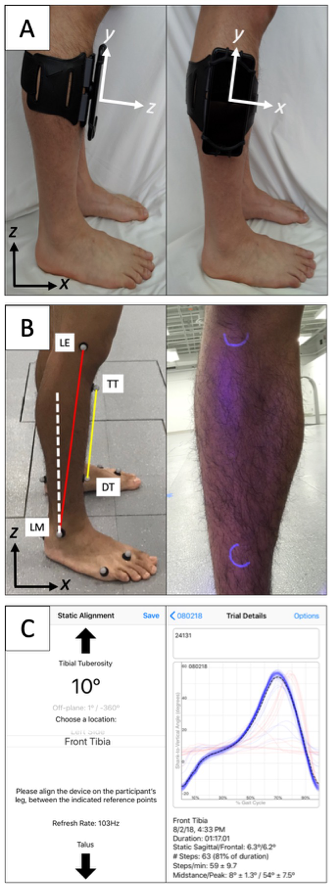
Method

Four licensed clinicians (two physical therapists and two orthotists) measured SVA during gait with a smartphone attached to the anterior or lateral shank surface of unimpaired adults.  We compared SVA calculated from the smartphone’s inertial measurement unit to marker-based measurements. Each clinician completed three sessions/day on two days with each participant to assess repeatability.

Results

Average absolute differences in SVA measured with a smartphone versus marker-based 3D motion analysis during gait were 0.67 ± 0.25° and 4.89 ± 0.72°, with anterior or lateral smartphone positions, respectively. The inter- and intra-day repeatability of SVA were within 2° for both smartphone positions.

Conclusions

Smartphone sensors can be used to measure SVA with high accuracy and repeatability during unimpaired gait, providing a widely-available tool for quantitative gait assessments.

Try it out!

The app for monitoring shank-to-vertical angle is available for you to download and use on either Android or iOS smartphone. Please complete THIS SURVEY which will then send you an e-mail with instructions for installation and use. This app is not an FDA approved medical device and should be used appropriately.