BR Shuman, MH Schwartz, KM Steele (2017) “Electromyography Data Processing Impacts Muscle Synergies during Gait for Unimpaired Children and Children with Cerebral Palsy.” Frontiers in Computational Neuroscience

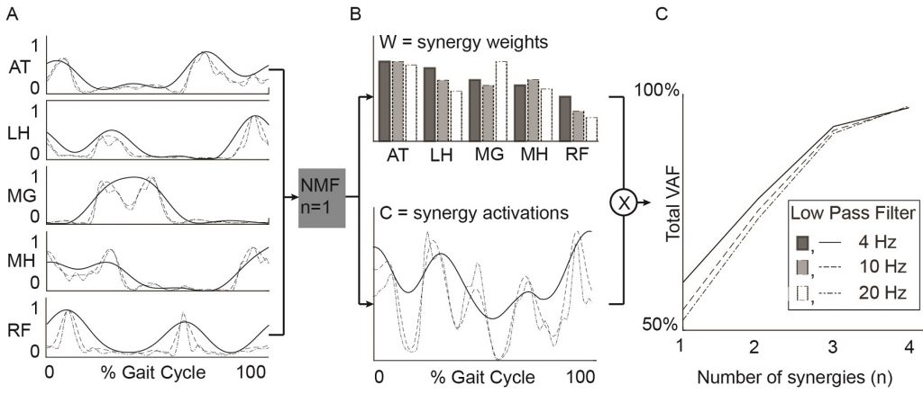
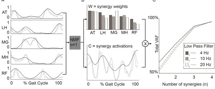
Example data from a representative TD participant. Left: EMG data processed with varying LP filter cutoffs. Center: Synergy weights and activations. Right: Total variance accounted for by n synergies. Total variance accounted for by a given number of synergies was sensitive to LP filter choice and decreased in both TD and CP groups with increasing LP cutoff frequency.

Journal article in Frontiers in Computational Neuroscience:

Filtering parameters impact the results from muscle synergy analyses.

AExample data from a representative TD participant. Left: EMG data processed with varying LP filter cutoffs. Center: Synergy weights and activations. Right: Total variance accounted for by n synergies. Total variance accounted for by a given number of synergies was sensitive to LP filter choice and decreased in both TD and CP groups with increasing LP cutoff frequency.bstract: Muscle synergies calculated from electromyography (EMG) data identify weighted groups of muscles activated together during functional tasks. Research has shown that fewer synergies are required to describe EMG data of individuals with neurologic impairments. When considering potential clinical applications of synergies, understanding how EMG data processing impacts results and clinical interpretation is important. The aim of this study was to evaluate how EMG signal processing impacts synergy outputs during gait. We evaluated the impacts of two common processing steps for synergy analyses: low pass (LP) filtering and unit variance scaling. We evaluated EMG data collected during barefoot walking from five muscles of 113 children with cerebral palsy (CP) and 73 typically-developing (TD) children. We applied LP filters to the EMG data with cutoff frequencies ranging from 4 to 40 Hz (reflecting the range reported in prior synergy research). We also evaluated the impact of normalizing EMG amplitude by unit variance. We found that the total variance accounted for (tVAF) by a given number of synergies was sensitive to LP filter choice and decreased in both TD and CP groups with increasing LP cutoff frequency (e.g., 9.3 percentage points change for one synergy between 4 and 40 Hz). This change in tVAF can alter the number of synergies selected for further analyses. Normalizing tVAF to a z-score (e.g., dynamic motor control index during walking, walk-DMC) reduced sensitivity to LP cutoff. Unit variance scaling caused comparatively small changes in tVAF. Synergy weights and activations were impacted less than tVAF by LP filter choice and unit variance normalization. These results demonstrate that EMG signal processing methods impact outputs of synergy analysis and z-score based measures can assist in reporting and comparing results across studies and clinical centers.
KM Steele, RW Jackson, BR Shuman, SH Collins (2017) “Muscle recruitment and coordination with an ankle exoskeleton.” Journal of Biomechanics

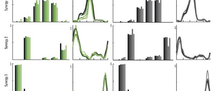
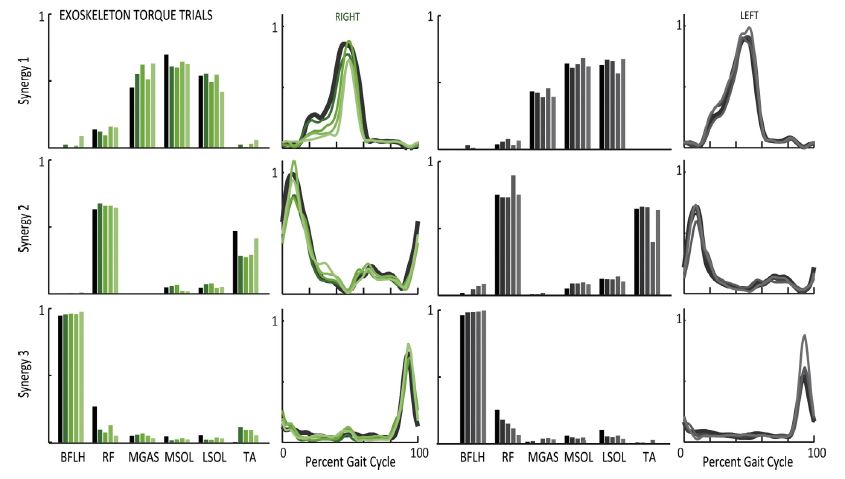
Synergy structure and activations had minimal changes with increasing exoskeleton torque.

Journal article in Journal of Biomechanics:

How do muscle activations and synergies change when an individual wears an ankle exoskeleton during gait?

Abstract: Exoskeletons have the potential to assist and augment human performance. Understanding how users adapt their movement and neuromuscular control in response to external assistance is important to inform the design of these devices. The aim of this research was to evaluate changes in muscle recruitment and coordination for ten unimpaired individuals walking with an ankle exoskeleton. We evaluated changes in the activity of individual muscles, cocontraction levels, and synergistic patterns of muscle coordination with increasing exoskeleton work and torque. Participants were able to selectively reduce activity of the ankle plantarflexors with increasing exoskeleton assistance. Increasing exoskeleton net work resulted in greater reductions in muscle activity than increasing exoskeleton torque. Patterns of muscle coordination were not restricted or constrained to synergistic patterns observed during unassisted walking. While three synergies could describe nearly 95% of the variance in electromyography data during unassisted walking, these same synergies could describe only 85–90% of the variance in muscle activity while walking with the exoskeleton. Synergies calculated with the exoskeleton demonstrated greater changes in synergy weights with increasing exoskeleton work versus greater changes in synergy activations with increasing exoskeleton torque. These results support the theory that unimpaired individuals do not exclusively use central pattern generators or other low-level building blocks to coordinate muscle activity, especially when learning a new task or adapting to external assistance, and demonstrate the potential for using exoskeletons to modulate muscle recruitment and coordination patterns for rehabilitation or performance.Synergy structure and activations had minimal changes with increasing exoskeleton torque.

NIH cerebral palsy strategic plan – our comments

The National Institutes of Health recently released the “Strategic Plan for Cerebral Palsy Research” which outlines challenges and priorities to guide future research to improve the lives of people with cerebral palsy.

Our diverse research group enjoyed reading and discussing this plan, which will likely influence our future research goals and support. We’ve shared our group’s comments, organized and prepared by Dr. Heather Feldner, below:

“Our research group appreciated the committee’s focus on creating a centralized data source for CP, attention to the needs and perspectives of adults with CP, their childhood experiences, and their transition from pediatric to adult healthcare providers, and the call for greater caregiver support services and patient-reported outcomes. However, we also had concerns. First, the terminology is inconsistent and often inappropriate. “Cure”, “damage”, and the implication that people with CP cannot be “healthy” is not empowering language in supporting the lives, unique contributions, and perspectives of people with CP as diverse and valued individuals in our society. Further, while advocates of people with CP were included in this stakeholder group, there is a concerning lack of people who actually have a diagnosis of CP, when these should be the primary stakeholders setting a research agenda about their own lives and needs. Finally, given the uncertainty of government funding agencies like the NIH under the current administration’s budget proposal, and the speed of science of translating research from bench to bedside, it appears that too little priority has been placed on interventions or programs that could have an influence right now for the people living with CP in the US dealing with self-identified participation issues such as access to employment and education, as well as impairment-related needs such as pain management, access to technology, and functional mobility.

We are excited that NIH is engaged to set a national research agenda for cerebral palsy and we look forward to continuing to serve this community.

Logo of NINDS/NICHD Plan for cerebral palsy research

B Soran, L Lowes, KM Steele (2016) “Evaluation of infants with spinal muscular atrophy using convolutional neural networks.” European Conference on Computer Vision

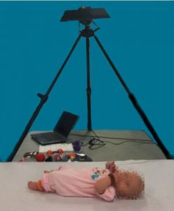
Experimental set-up with infant positioned below Kinect depth camera.

Peer-reviewed paper at European Conference on Computer Vision:

30-second videos from a depth camera can be used in the evaluation of infants with spinal muscular atrophy.

Experimental set-up with infant positioned below Kinect depth camera.Abstract: Spinal Muscular Atrophy is the most common genetic cause of infant death. Due to its severity, there is a need for methods for automated estimation of disease progression. In this paper we propose a Convolutional-Neural-Network (CNN) model to estimate disease progression during infants’ natural behavior. With the proposed methodology, we were able to predict each child’s score on current behavior-based clinical exams with an average per-subject error of 6.96 out of 72 points (<10 % difference), using 30-second videos in leave-one-subject-out-cross-validation setting. When simple statistics were used over 30-second video-segments to estimate a score for longer videos, we obtained an average error of 5.95 (8 % error rate). By showing promising results on a small dataset (N = 70, 2-minute samples, which were handled as 1487, 30-second video segments), our methodology demonstrates that it is possible to benefit from CNNs on small datasets by proper design and data handling choices.

J Wu, BR Shuman, BW Brunton, KM Steele, JD Olson, RPN Rao (2016) “Multistep model for predicting upper-limb 3D isometric force application from pre-movement electrocorticographic features.” IEEE Engineering Medicine & Biology

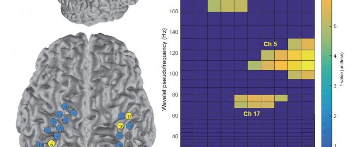
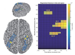
Example of ECoG recording during upper-extremity force production.

Peer-reviewed paper at IEEE Engineering in Medicine & Biology Annual Conference:

Can we estimate upper-extremity force production from electrocorticographic recordings?

Example of ECoG recording during upper-extremity force production.Abstract: Neural correlates of movement planning onset and direction may be present in human electrocorticography in the signal dynamics of both motor and non-motor cortical regions. We use a three-stage model of jPCA reduced-rank hidden Markov model (jPCA-RR-HMM), regularized shrunken-centroid discriminant analysis (RDA), and LASSO regression to extract direction-sensitive planning information and movement onset in an upper-limb 3D isometric force task in a human subject. This mode achieves a relatively high true positive force-onset prediction rate of 60% within 250ms, and an above-chance 36% accuracy (17% chance) in predicting one of six planned 3D directions of isometric force using pre-movement signals. We also find direction-distinguishing information up to 400ms before force onset in the pre-movement signals, captured by electrodes placed over the limb-ipsilateral dorsal premotor regions. This approach can contribute to more accurate decoding of higher-level movement goals, at earlier timescales, and inform sensor placement. Our results also contribute to further understanding of the spatiotemporal features of human motor planning.