KM Peters, VE Kelly, T Chang, MC Weismann, S Westcott McCoy, KM Steele (2018) “Muscle recruitment and coordination during upper-extremity functional tests.” Journal of Electromyography and Kinesiology

Journal article in Journal of Electromyography and Kinesiology:

In collaboration with Rehabilitation Medicine here at the University of Washington, we evaluated muscle use of 20 unimpaired participants during three upper-extremity functional tests. An interactive supplement can be found HERE.
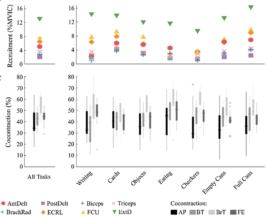
Recruitment and cocontration plots of eight upper-extremity muscles during the Jebsen Taylor Hand Function Test.Background: Performance-based tests, such as the Jebsen Taylor Hand Function Test or Chedoke Arm and Hand Activity Inventory, are commonly used to assess functional performance after neurologic injury. However, the muscle activity required to execute these tasks is not well understood, even for unimpaired individuals. The purpose of this study was to evaluate unimpaired muscle recruitment and coordination of the dominant and non-dominant limbs during common clinical tests.

Methods: Electromyography (EMG) recordings from eight arm muscles were monitored bilaterally for twenty unimpaired participants while completing these tests. Average signal magnitudes, activation times, and cocontraction levels were calculated from the filtered EMG data, normalized by maximum voluntary isometric contractions (MVICs).

Results: Overall, performance of these functional tests required low levels of muscle activity, with average EMG magnitudes less than 6.5% MVIC for all tests and muscles, except the extensor digitorum, which had higher activations across all tasks (11.7 ± 2.7% MVIC, dominant arm). When averaged across participants, cocontraction was between 25 and 62% for all tests and muscle pairs.

Conclusion: Tasks evaluated by speed of completion, rather than functional quality of movement demonstrated higher levels of muscle recruitment. These results provide baseline measurements that can be used to evaluate muscle-specific deficits after neurologic injury and track recovery using common clinical tests.



Daniel Ballesteros, Wing-Sum Law, and Claire Mitchell Present

Congratulations to Daniel, Wing-Sum and Claire for their excellent work this summer. We had the privilege of hosting three undergraduate students through the Summer Scholars program through Co-Motion and the Center for Sensorimotor Neural Engineering here at the University of Washington.

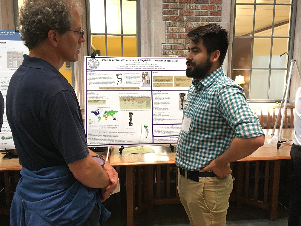
Daniel presented in Mary Gates Hall on his research involving the implementation of a pediatric exoskeleton into low-resource countries:

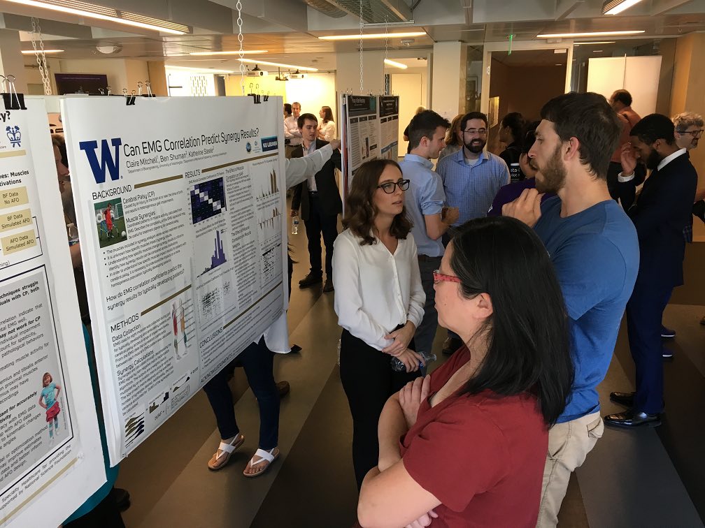
Daniel fields a question from a member of the University of Washington community during his poster session at Mary Gates Hall. Claire researched how varying muscles impact synergy outcomes:


Claire Mitchell presents her poster at the Center for Sensorimotor Neural Engineering


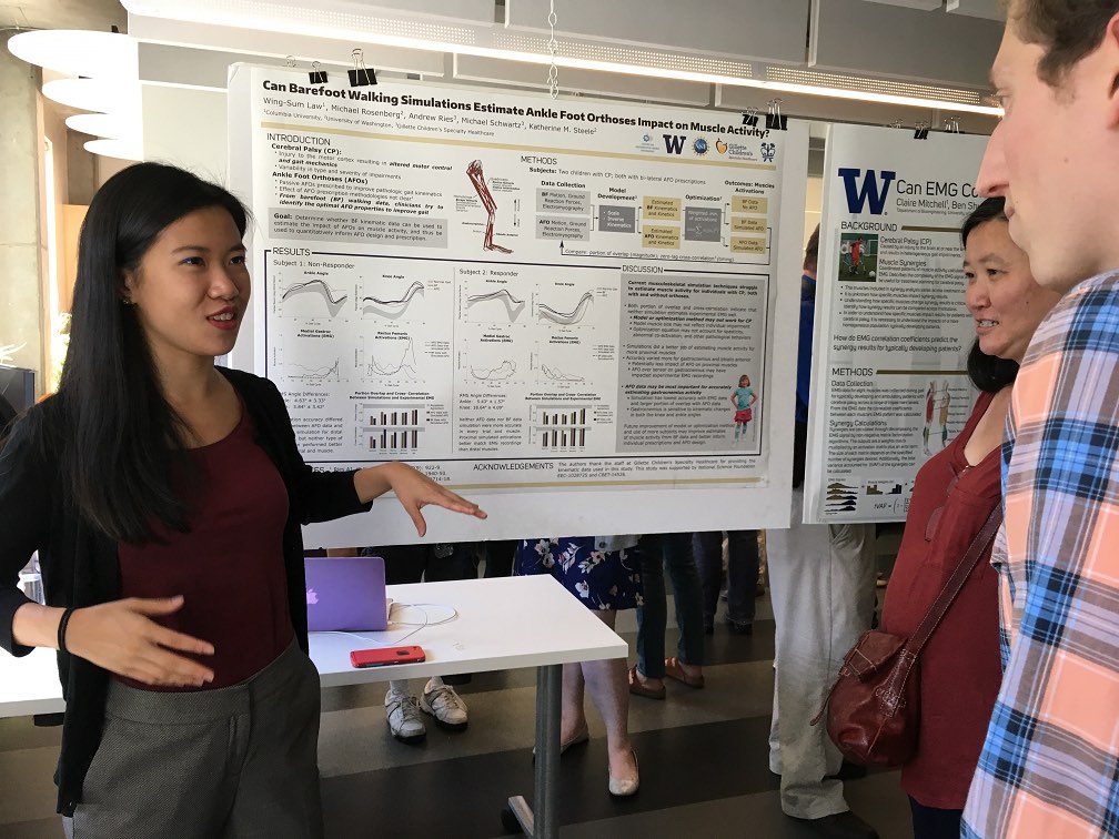
Wing-Sum assessed the impact of an ankle foot orthosis on muscle demands in children with cerebral palsy and typically developing peers:

Wing-Sum presents her research during a poster session

Lab members present at the 2017 American Society of Biomechanics

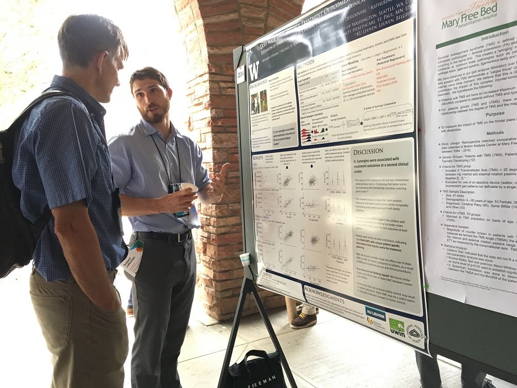
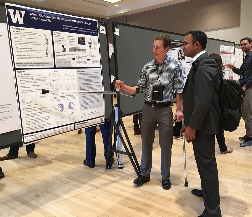
Ben Shuman, Michael Rosenberg, Keshia Peters, and Kat Steele all presented posters during the 2017 American Society of Biomechanics conference in Boulder, CO.

Dr. Steele also presented during a session which encompassed ASB and the Gait and Clinical Movement Analysis Society to invoke discussion about technical challenges in clinical motion analysis.

Great work, everyone!

Ben Shuman discusses his poster Michael Rosenberg walks through the results section of his poster with a fellow ASB member.

M Rosenberg, KM Steele (2017) “Simulated impacts of ankle foot orthoses on muscle demand and recruitment in typically-developing children and children with cerebral palsy and crouch gait.” PLoS ONE

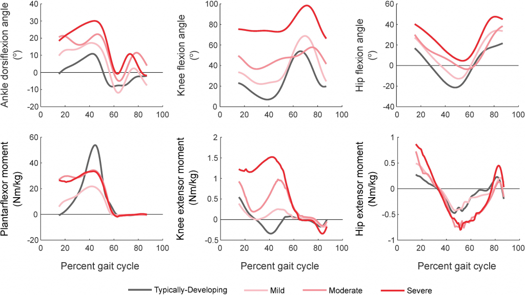
Top: Ankle, knee and hip kinematics for gait in TD children and children with crouch gait. TD children walked with less ankle dorsiflexion and knee flexion during stance than those with crouch gait. Bottom: Ankle, knee and hip moments for gait in TD children and crouch gait. TD children generated larger peak plantarflexor moments and smaller peak knee extensor moments compared to crouch gait. Knee extensor moments increased with increasing crouch severity.

Journal article in PLOS ONE:

Michael Rosenberg and Kat Steele investigate the impacts of ankle foot orthoses on children with cerebral palsy and typically-developing peers through simulation.

Background

Passive ankle foot orthoses (AFOs) are often prescribed for children with cerebral palsy (CP) to assist locomotion, but predicting how specific device designs will impact energetic demand during gait remains challenging. Powered AFOs have been shown to reduce energy costs of walking in unimpaired adults more than passive AFOs, but have not been tested in children with CP.

Aim

The goal of this study was to investigate the potential impact of powered and passive AFOs on muscle demand and recruitment in children with CP and crouch gait.

Method

We simulated gait for nine children with crouch gait and three typically-developing children with powered and passive AFOs. For each AFO design, we computed reductions in muscle demand compared to unassisted gait.

Results

Powered AFOs reduced muscle demand 15–44% compared to unassisted walking, 1–14% more than passive AFOs. A slower walking speed was associated with smaller reductions in absolute muscle demand for all AFOs (r2 = 0.60–0.70). However, reductions in muscle demand were only moderately correlated with crouch severity (r2 = 0.40–0.43). The ankle plantarflexor muscles were most heavily impacted by the AFOs, with gastrocnemius recruitment decreasing 13–73% and correlating with increasing knee flexor moments (r2 = 0.29–0.91).

Interpretation

These findings support the potential use of powered AFOs for children with crouch gait, and highlight how subject-specific kinematics and kinetics may influence muscle demand and recruitment to inform AFO design. PDF

Top: Ankle, knee and hip kinematics for gait in TD children and children with crouch gait. TD children walked with less ankle dorsiflexion and knee flexion during stance than those with crouch gait. Bottom: Ankle, knee and hip moments for gait in TD children and crouch gait. TD children generated larger peak plantarflexor moments and smaller peak knee extensor moments compared to crouch gait. Knee extensor moments increased with increasing crouch severity.