 “Dynamic motor control is associated with treatment outcomes for children with cerebral palsy.” Developmental Medicine & Child Neurology")
Journal article in Developmental Medicine & Child Neurology:
Kat Steele partnered with Michael Schwartz from Gillette Children’s Specialty Healthcare to investigate the impact of dynamic motor control on varying treatments in children with cerebral palsy.
Aim
To estimate the impact of dynamic motor control on treatment outcomes in children with cerebral palsy.
Method
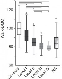
We used multiple regression on a retrospective cohort of 473 ambulatory children with cerebral palsy who underwent conservative treatment, single-level orthopaedic surgery, single-event multi-level orthopaedic surgery, or selective dorsal rhizotomy. Outcomes included gait pattern, gait speed, energy cost of walking, and the Pediatric Outcomes Data Collection Instrument. Explanatory variables considered were pre-treatment levels of each outcome, treatment group, prior treatment, age, and dynamic motor control computed from surface electromyography using synergy analysis. Effect sizes were estimated from the adjusted response.
Results
Pre-treatment levels had effect sizes 2 to 13 times larger than the next largest variable. Individuals with milder pre-treatment involvement had smaller gains or actual declines. Dynamic motor control was significant in all domains except energy cost. The effect size of dynamic motor control was second only to pre-treatment level, and was substantially larger than the effect size of treatment group for outcomes where both were significant (gait pattern 2:1, gait speed 4:1). The effect of dynamic motor control was independent of treatment group.
Interpretation
Dynamic motor control is an important factor in treatment outcomes. Better dynamic motor control is associated with better outcomes, regardless of treatment. PDF
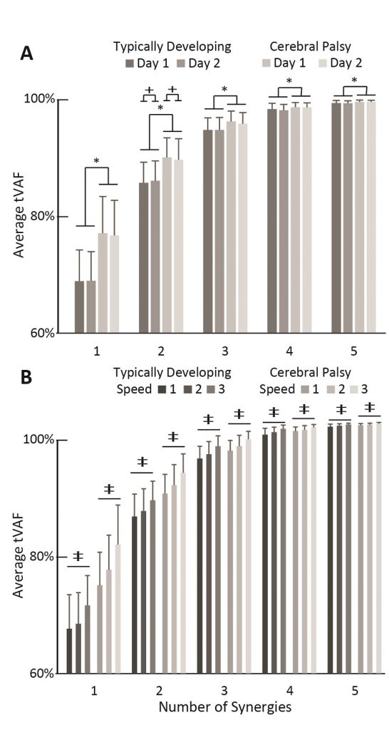
 “Repeatability of muscle synergies within and between days for typically developing children and children with cerebral palsy.” Gait & Posture.")
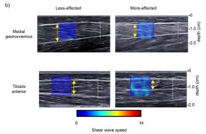
 “Use of shear wave ultrasound elastography to quantify muscle properties in cerebral palsy.” Clinical Biomechanics")
 “Muscle synergies and complexity of neuromuscular control during gait in cerebral palsy.” Developmental Medicine & Child Neurology")
 “Including universal design in engineering courses to attract diverse students.” Proceedings of the American Society for Engineering Education")