BR Shuman, M Goudriaan, K Desloovere, MH Schwartz, KM Steele (2018) “Associations Between Muscle Synergies and Treatment Outcomes in Cerebral Palsy Are Robust Across Clinical Centers.” Archives of Physical Medicine and Rehabilitation

Journal article in Archives of Physical Medicine and Rehabilitation:

In collaboration with Gillette Children’s Hospital and University Hospital Pellenberg we examined whether associations between treatment outcomes and muscles synergies are robust between clinical centers.

Objective: To determine whether patient-specific differences in motor control quantified using muscle synergy analysis were associated with changes in gait after treatment of cerebral palsy (CP) across 2 clinical centers with different treatments and clinical protocols.
Design: Retrospective cohort study.
Setting: Clinical medical center.
Participants: Center 1: children with CP (n=473) and typically developing (TD) children (n=84). Center 2: children with CP (n=163) and TD children (n=12).
Interventions: Standard clinical care at each center.
Main outcome measures: The Dynamic Motor Control Index During Walking (walk-DMC) was computed from electromyographic data during gait using muscle synergy analysis. Regression analysis was used to evaluate whether pretreatment walking speed or kinematics, muscle synergies, treatment group, prior treatment, or age were associated with posttreatment changes in gait at both clinical centers.
Results: Walk-DMC was significantly associated with changes in speed and kinematics after treatment with similar regression models at both centers. Children with less impaired motor control were more likely to have improvements in walking speed and gait kinematics after treatment, independent of treatment group.
Conclusions: Dynamic motor control evaluated with synergy analysis was associated with changes in gait after treatment at both centers, despite differences in treatments and clinical protocols. This study further supports the finding that walk-DMC provides additional information, not captured in traditional gait analysis, that may be useful for treatment planning.

Alyssa Spomer and Momona Yamagami Present at a Neurorehabilitation Conference in Spain

Alyssa and Momona attended the Summer School on Neurorehabilitation (SSNR) in Baiona, Spain from September 16th to the 21st. Alyssa gave a podium presentation on a feedback system she is developing that aims to characterize and target altered motor control in cerebral palsy. Momona gave a poster presentation to share her recent quantifications of deficits in motor planning in cerebral palsy. Nice work, Alyssa and Momona!Alyssa at podium presenting "A Feedback System to Characterize and Target Altered Motor Control in Cerebral Palsy".


Monoma stands in front of her poster "Quantification of Deficits in Motor Planning in Cerebral Palsy", discussing with interested colleagues.


Brianna Goodwin Presents at Seattle Children’s Grand Rounds

Brianna Goodwin, a Master’s student in our lab, presented her collaborative Brianna Goodwin is pictured behind a podium, standing in front of her slideshow during her presentation.abstract on monitoring Constraint-Induced Movement Therapy (CIMT), a therapy for children with hemiplegic cerebral palsy (CP), at the Seattle Children’s Hospital (SCH) Grand Rounds this past week. The Grand Rounds are a time to present research, new ideas, and translational science to medical personnel of varied background.

To read Brianna’s abstract in full, download her PDF here: SCH Grand Rounds, CIMT abstract


M Goudriaan, BR Shuman, KM Steele, M Van den Hauwe, N Goemans, G Molenaers, K Desloovere (2018) “Non-neural Muscle Weakness Has Limited Influence on Complexity of Motor Control during Gait.” Frontiers in Human Neuroscience

Journal Article in Frontiers in Human Neuroscience:

Despite significant differences in kinematics children with Duchenne muscular dystrophy have similar control complexity to typically developing children.

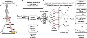
Abstract: Cerebral palsy (CP) and Duchenne muscular dystrophy (DMD) are neuromuscular disorders characterized by muscle weakness. Weakness in CP has neural and non-neural components, whereas in DMD, weakness can be considered as a predominantly non-neural problem. Despite the different underlying causes, weakness is a constraint for the central nervous system when controlling gait. CP demonstrates decreased complexity of motor control during gait from muscle synergy analysis, which is reflected by a higher total variance accounted for by one synergy (tVAF1). However, it remains unclear if weakness directly contributes to higher tVAF1 in CP, or whether altered tVAF1 reflects mainly neural impairments. If muscle weakness directly contributes to higher tVAF1, then tVAF1 should also be increased in DMD. To examine the etiology of increased tVAF1, muscle activity data of gluteus medius, rectus femoris, medial hamstrings, medial gastrocnemius, and tibialis anterior were measured at self-selected walking speed, and strength data from knee extensors, knee flexors, dorsiflexors and plantar flexors, were analyzed in 15 children with CP [median (IQR) age: 8.9 (2.2)], 15 boys with DMD [8.7 (3.1)], and 15 typical developing (TD) children [8.6 (2.7)]. We computed tVAF1 from 10 concatenated steps with non-negative matrix factorization, and compared tVAF1between the three groups with a Mann-Whiney U-test. Spearman’s rank correlation coefficients were used to determine if weakness in specific muscle groups contributed to altered tVAF1. No significant differences in tVAF1 were found between DMD [tVAF1: 0.60 (0.07)] and TD children [0.65 (0.07)], while tVAF1 was significantly higher in CP [(0.74 (0.09)] than in the other groups (both p < 0.005). In CP, weakness in the plantar flexors was related to higher tVAF1 (r = −0.72). In DMD, knee extensor weakness related to increased tVAF1 (r = −0.50). These results suggest that the non-neural weakness in DMD had limited influence on complexity of motor control during gait and that the higher tVAF1 in children with CP is mainly related to neural impairments caused by the brain lesion.

H Choi, KM Peters, M MacConnell, K Ly, E Eckert, KM Steele (2017) “Impact of ankle foot orthosis stiffness on Achilles tendon and gastrocnemius function during unimpaired gait.” Journal of Biomechanics

Journal article in Journal of Biomechanics:

How does the stiffness of an AFO impact the muscultendon dynamics of the gastrocnemius?

Abstract

Method combining ultrasound and musculoskeletal modeling to evaluate changes in muscle and tendon length.

Ankle foot orthoses (AFOs) are designed to improve gait for individuals with neuromuscular conditions and have also been used to reduce energy costs of walking for unimpaired individuals. AFOs influence joint motion and metabolic cost, but how they impact muscle function remains unclear. This study investigated the impact of different stiffness ankle foot orthoses (AFOs) on medial gastrocnemius muscle (MG) and Achilles tendon (AT) function during two different walking speeds. We performed gait analyses for eight unimpaired individuals. Each individual walked at slow and very slow speeds with a 3D printed AFO with no resistance (free hinge condition) and four levels of ankle dorsiflexion stiffness: 0.25 Nm / °, 1 Nm / °, 2 Nm / °, and 3.7 Nm / °. Motion capture, ultrasound, and musculoskeletal modeling were used to quantify MG and AT lengths with each AFO condition. Increasing AFO stiffness increased peak AFO dorsiflexion moment with decreased peak knee extension and peak ankle dorsiflexion angles. Overall musculotendon length and peak AT length decreased, while peak MG length increased with increasing AFO stiffness. Peak MG activity, length, and velocity significantly decreased with slower walking speed. This study provides experimental evidence of the impact of AFO stiffness and walking speed on joint kinematics and musculotendon function. These methods can provide insight to improve AFO designs and optimize musculotendon function for rehabilitation, performance, or other goals.