 Congratulations to Michael Rosenberg for being awarded the Komor New Investigator Award at the 2021 Technical Group on Computer Simulation (TGCS) at ISB. This award is presented at each symposium to recognize the best paper by a new investigator. Way to go Michael!
Projects
Congratulations, Alyssa! Alyssa Spomer passes her Ph.D. General Exam
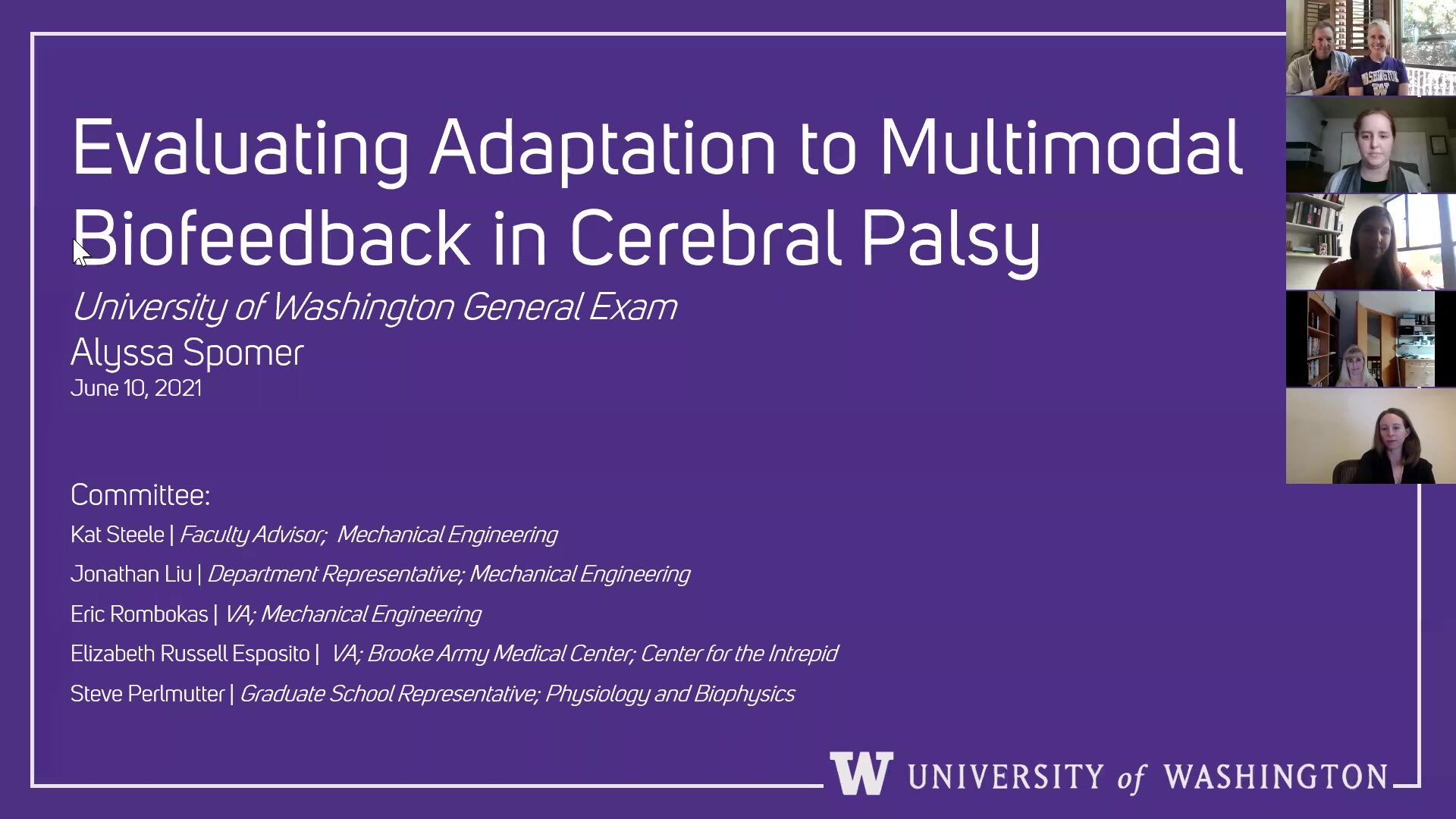 Congratulations to Alyssa Spomer for passing her general exam!
Alyssa’s proposed work titled Evaluating Adaptation to Multimodal Biofeedback in Cerebral Palsy was approved by her Ph.D. committee. Way to go Alyssa!
Momona Yamagami wins the College of Engineering Student Research Award. Congratulations Momona!
 The College of Engineering Awards acknowledges the extraordinary efforts of the college’s teaching and research assistants, staff, and faculty members. Momona Yamagami was selected for the 2021 Student Research Award. Congratulations Momona!
Momona Yamagami is an innovative researcher who focuses on developing novel accessible technologies with translational impact. In her first year, she helped build an interdisciplinary research program that blended neuroengineering, human-computer interaction and rehabilitation at the Amplifying Motion and Performance (AMP) Lab to evaluate and mitigate symptoms of Parkinson’s disease using virtual reality. Dedicated to building accessible and inclusive technology, she is working to apply control theory and artificial intelligence to improve device accessibility for people with and without limited motion.
“Momona is a truly exceptional student with a demonstrated history of leadership in research and education. We cannot wait to see where Momona steers her career trajectory and research contributions.”
NSF Convergence Accelerator | Living Better through Rehabilitation & Assistive Technology
A second NSF Convergence Accelerator focused on increasing access and inclusion. The LIBERATE workshop is focused on Living Better through Rehabilitation & Assistive Technology.
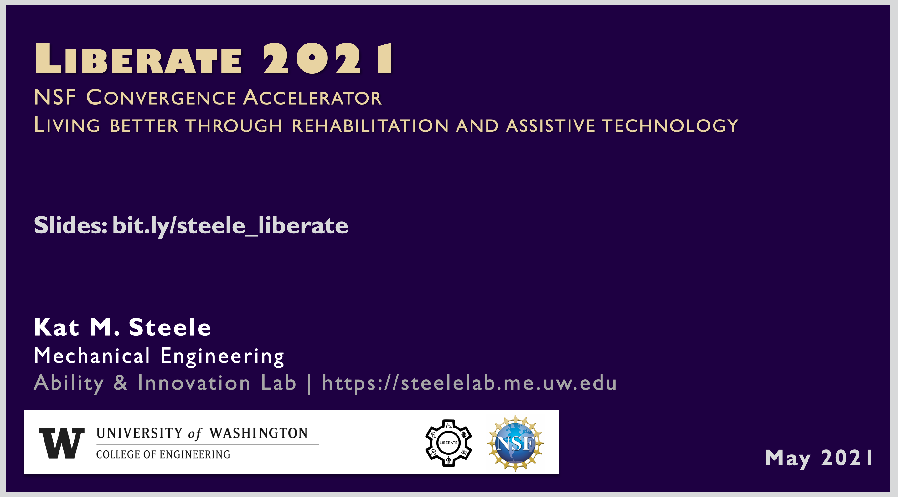
As an NSF Convergence Accelerator, participants will seek to identify pathways that could be pursued by multidisciplinary teams to get solutions at least to a prototype stage in 3-5 years. The long-term goal from this workshop is to kickstart the next wave of technologies that will empower people with disabilities.
Dr. Steele will be participating and presenting some kernels of ideas for inclusion, especially highlighting recent work from CREATE.
Slides
Email Dr. Steele (kmsteele – at – uw – dot – edu) with questions, comments, or suggestions.
NSF Convergence Accelerator | Inclusion in the Workplace
The NSF Convergence Accelerator on Accelerating Disability Inclusion in Workplaces through Technology starts on May 20th.
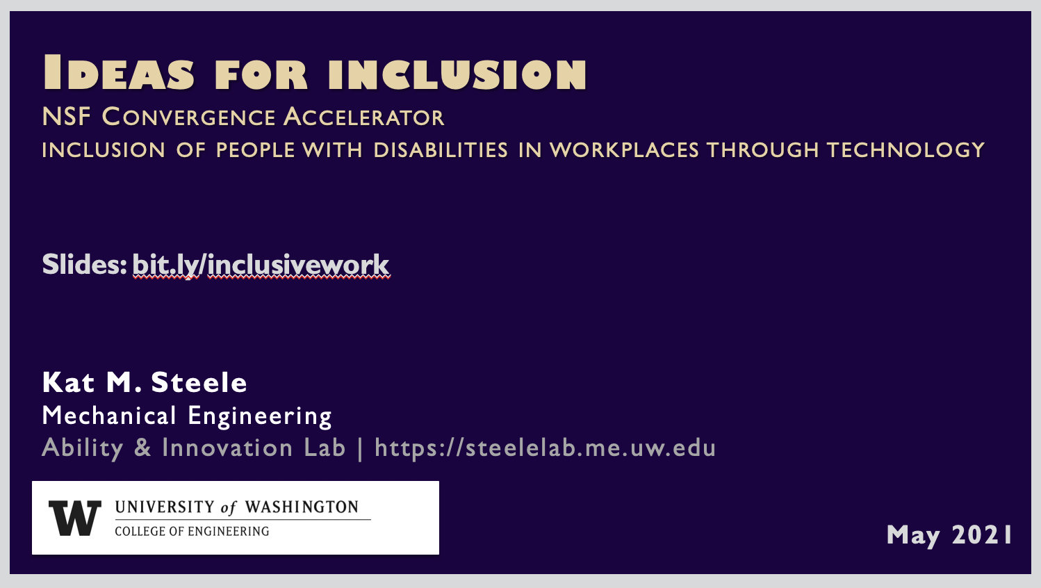
The goals for this workshop are to identify pathways for technology to solve or mitigate accessibility and inclusion challenges in current and emerging workplaces. As an NSF Convergence Accelerator, participants will seek to identify pathways that could be pursued by multidisciplinary teams to get solutions at least to a prototype stage in 3-5 years. The long-term goals from this workshop are to set in motion paradigm shifts that brings the percentage of individuals with disabilities participating in the workforce closer to the general population.
Dr. Steele will be presenting some ideas on inclusion in the workplace – from work environments to transportation to workforce development.
Slides
Email Dr. Steele (kmsteele – at – uw – dot – edu) with questions, comments, or suggestions.