HA Feldner, C Papazian, KM Peters, CJ Cruetzfeldt, KM Steele (2021) “Clinical Use of Surface Electromyography to Track Acute Upper Extremity Muscle Recovery after Stroke: A Descriptive Case Study of a Single Patient”

Journal Article in Applied System Innovation:

This work highlights the potential of wearable technologoies to monitor muscle activity changes during stroke recovery in acute clinical settings and their importance for motivation and understanding of progression from the survivor’s point of view: ‘I was hopeful that it would show signs of things that are occurring when I couldn’t physically feel it…if you had other scientific evidence that things were happening, even beyond their notion that it would, it gives you a lot of hope. You just have to be patient, and it’s harder to take when someone tells you, but easier to understand if someone actually shows you’.

Left image depicts arm with pads placed over muscle with right pictures depicting similar image

Aim: Describe the use of wireless sEMG sensors to examine changes in muscle activity during acute and subacute phases of stroke recovery, and understand the participant’s perceptions of sEMG monitoring.

Method: Muscle activity was tracked by five wireless sEMG sensors beginning three days post-stroke and continued through discharge from inpatient rehabilitation. Activity logs were completed each session, and a semi-structured interview occurred at the final session with three- and eight-month follow-up sessions.

Results: The longitudinal monitoring of muscle and movement recovery in the clinic and community was feasible using sEMG sensors. The participant and medical team felt monitoring was unobtrusive, interesting, and motivating for recovery, but desired greater in-session feedback to inform rehabilitation.

Interpretation: This work highlights that barriers in equipment and signal quality still exist, but capitalizing on wearable sensing technology in the clinic holds promise for enabling personalized stroke recovery.

MC Rosenberg, BS Banjanin, SA Burden, KM Steele (2020) “Predicting walking response to ankle exoskeleton using data driven models”

Journal Article in The Royal Society:

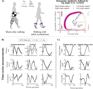
This work highlights the potential of data-driven models grounded in dynamical systems theory to predict complex individualized responses to ankle exoskeletons., without requiring explicit knowledge of the individual’s physiology or motor control

silhouette walking on left with purple lines and projections on right elipsoids and colored spheres

Aim: Evaluate the ability of three classes of subject-specific phase-varying (PV) models to predict kinematic and myoelectric responses to ankle exoskeletons during walking, without requiring prior knowledge of specific user characteristics.

Method: Data from 12 unimpaired adults walking with bilateral passive ankle exoskeletons were captured. PV, linear PV (LPV), and nonlinear PV (NPV) models leveraged Floquet theory to kinematics and muscle activity in response to three exoskeleton torque conditions.

Results: The LPV model’s predictions were more accurate than the PV model when predicting less than 12.5% of a stride in the future and explained 49–70% of the variance in hip, knee and ankle kinematic responses to torque. The LPV model also predicted kinematic responses with similar accuracy to the more-complex NPV model. Myoelectric responses were challenging to predict with all models, explaining at most 10% of the variance in responses.

Interpretation: This work highlights the potential of data-driven PV models to predict complex subject-specific responses to ankle exoskeletons and inform device design and control.

M Yamagami, KM Steele, SA Burden (2020) “Decoding Intent With Control Theory: Comparing Muscle Versus Manual Interface Performance”

Journal Article in ACM Conference on Human Factors in Computing Systems (CHI) 2020 Preceedings:

These results suggest that control theory modeling can provide a platform to successfully quantify device performance in the absence of errors arising from motor impairments

Split image of upper body of user holding rod and slider with computer screen

Photo (top and bottom) of a user using a slider (top) and muscles (bottom) to control a cursor on the screen.
(Top image) Side image of user. User rests their elbow and pinches the slider and moves the slider towards and away from their body to control the cursor.
(Bottom image) Side image of user. User is strapped to a rigid device holding a bar with hands supinated towards the ceiling, with the forearms at a 90 degree angle from the upper arms.
Electrodes are placed on the biceps and triceps and labelled. Arrows pointing up and down indicate that users move their arm up and down to control the cursor.


Background: Manual device interaction requires precise coordination which may be difficult for users with motor impairments. Muscle interfaces provide alternative interaction methods that may enhance performance, but have not yet been evaluated for simple (eg. mouse tracking) and complex (eg. driving) continuous tasks. Control theory enables us to probe continuous task performance by separating user input into intent and error correction to quantify how motor impairments impact device interaction

Aim:  Propose and extend an experimental and analytical method to guide future development of accessible interfaces like muscle interfaces using control theory

Method: We compared the effectiveness of a manual versus a muscle interface for eleven users without and three users with motor impairments performing continuous tasks.

Results: Both user groups preferred and performed better with the muscle versus the manual interface for the complex continuous task.

Interpretation: Results suggest muscle interfaces and algorithms that can detect and augment user intent may be especially useful for future design of interfaces for continuous tasks.


Momona also gave a phenomenal talk on this paper last week in the University of Washington’s ‘DUB Shorts’ series (video posted below). Nice job Momona!

NL Zaino, KM Steele, JM Donelan, MH Schwartz (2020) “Energy consumption does not change after selective dorsal rhizotomy in children with spastic cerebral palsy” Developmental Medicine & Child Neurology

Journal Article in Developmental Medicine & Child Neurology:

This retrospective analysis demonstrated that energy consumption is not reduced after rhizotomy when compared to matched controls with cerebral palsy.

Spasticity and net-nondimensionalized (NN) energy consumption for children with cerebral palsy (CP) who underwent a selective dorsal rhizotomy (SDR) and matched peers with CP who did not undergo SDR (control). (a) Baseline spasticity and NN energy consumption were similar between groups. Gray lines show normative values for typically developing (TD) peers from Gillette Children’s Specialty Healthcare. (b) Spasticity and NN energy consumption decreased significantly at follow-up for both groups. The SDR cohort had a significantly greater decrease in spasticity compared to the no-SDR group, but a similar decrease in NN energy consumption. Bars represent distributions for each group including outliers (*).

Aim: To determine whether energy consumption changes after selective dorsal rhizotomy (SDR) among children with cerebral palsy (CP).

Method: We retrospectively evaluated net nondimensional energy consumption during walking among 101 children with bilateral spastic CP who underwent SDR (59 males, 42 females; median age [5th centile, 95th centile] 5y 8mo [4y 2mo, 9y 4mo]) compared to a control group of children with CP who did not undergo SDR. The control group was matched by baseline age, spasticity, and energy consumption (56 males, 45 females; median age [5th centile, 95th centile] 5y 8mo [4y 1mo, 9y 6mo]). Outcomes were compared at baseline and follow‐up (SDR: mean [SD] 1y 7mo [6mo], control: 1y 8mo [8mo]).

Results: The SDR group had significantly greater decreases in spasticity compared to matched controls (–42% SDR vs –20% control, p<0.001). While both groups had a modest reduction in energy consumption between visits (–12% SDR, –7% control), there was no difference in change in energy consumption (p=0.11) or walking speed (p=0.56) between groups.

Interpretation: The SDR group did not exhibit greater reductions in energy consumption compared to controls. The SDR group had significantly greater spasticity reduction, suggesting that spasticity had minimal impact on energy consumption during walking in CP. These results support prior findings that spasticity and energy consumption decrease with age in CP. Identifying matched control groups is critical for outcomes research involving children with CP to account for developmental changes.

B Nguyen, N Baicoianu, D Howell, KM Peters, KM Steele (2020) “Accuracy and repeatability of smartphone sensors for measuring shank-to-vertical angle” Prosthetics & Orthotics International

Journal Article in Prosthetics & Orthotics International

Example of how the smartphone app was used for this research. The top images show a black smartphone attached with a running arm band to the side or front of the shank - the two positions tested in this research. The middle figure shows the placement of the reflective markers for 3D motion analysis to evaluate the accuracy of the smartphone measurements. Markers were placed on the lateral epicondyle of the knee, lateral maleolus of the ankle, tibial tuberosity, and the distal tibia. Blacklight was used to mark the position of each marker and hide the position from the clinicians. The bottom panel shows screenshots from the app. The first screen is used to align the device and has arrows at the top and bottom that remind the clinician which anatomical landmarks should be used to align the device while displaying the shank-to-vertical angle in real time. The second screenshot shows an example of the calculated shank-to-vertical angle while someone was walking. The average is shown with a bold black line, with all other trials shown in blue and excluded trials (e.g., when someone was stopping or turning) that deviated more than one standard deviation from other trials are shown in red. There is also text below the graph that provides summary measures, like shank-to-vertical angle in mid stand and cadence (steps/min). The results can be exported as a picture or sent via e-mail using the app.
A) Smartphone positioning on the front or side of the shank. B) Reflective markers on the the tibial tuberosity (TT) – distal tibia (DT) and lateral epicondyle (LE) – lateral malleolus (LM) were used to compare the accuracy of the smartphone to traditional motion capture. UV markings were used to keep placement of these markers constant while blinding clinicians. C) Sample screenshots of the mobile application, including the set-up screen and results automatically produced after a walking trial.

Background

Assessments of human movement are clinically important. However, accurate measurements are often unavailable due to the need for expensive equipment or intensive processing. For orthotists and therapists, shank-to-vertical angle (SVA) is one critical measure used to assess gait and guide prescriptions. Smartphone-based sensors may provide a widely-available platform to expand access to quantitative assessments.

Objectives

Assess accuracy and repeatability of smartphone-based measurement of SVA compared to marker-based 3D motion analysis.

Method

Four licensed clinicians (two physical therapists and two orthotists) measured SVA during gait with a smartphone attached to the anterior or lateral shank surface of unimpaired adults.  We compared SVA calculated from the smartphone’s inertial measurement unit to marker-based measurements. Each clinician completed three sessions/day on two days with each participant to assess repeatability.

Results

Average absolute differences in SVA measured with a smartphone versus marker-based 3D motion analysis during gait were 0.67 ± 0.25° and 4.89 ± 0.72°, with anterior or lateral smartphone positions, respectively. The inter- and intra-day repeatability of SVA were within 2° for both smartphone positions.

Conclusions

Smartphone sensors can be used to measure SVA with high accuracy and repeatability during unimpaired gait, providing a widely-available tool for quantitative gait assessments.

Try it out!

The app for monitoring shank-to-vertical angle is available for you to download and use on either Android or iOS smartphone. Please complete THIS SURVEY which will then send you an e-mail with instructions for installation and use. This app is not an FDA approved medical device and should be used appropriately.