KM Steele, ME Munger, KM Peters, BR Shuman, MH Schwartz (2019) “Repeatability of electromyography recordings and muscle synergies during gait among children with cerebral palsy.” Gait & Posture

Journal Article in Gait & Posture:

Repeatability of EMG is similar between typically developing children and children with cerebral palsy.

Background: Clinical gait analysis is commonly used in the evaluation and treatment of children with cerebral palsy (CP). While the repeatability of kinematic and kinetic measures of gait has previously been evaluated, the repeatability of electromyography (EMG) recordings or measures calculated from EMG data, such as muscle synergies, remains unclear for this population.

Research Question: Are EMG recordings and muscle synergies from clinical gait analysis repeatable between visits for children with CP?

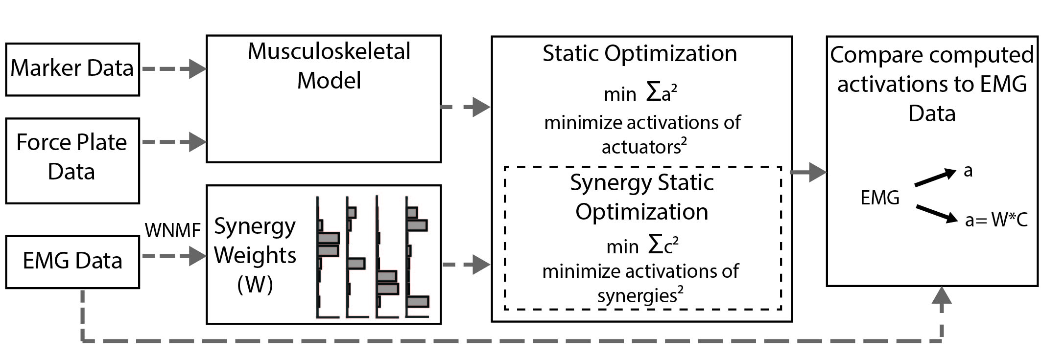
Methods: We recruited 20 children with bilateral CP who had been referred for clinical gait analysis. The children completed two visits less than six weeks apart with EMG data collected bilaterally from five muscles (rectus femoris, medial hamstrings, vastus lateralis, anterior tibialis, and medial gastrocnemius). Variance ratio and cosine similarity were used to evaluate repeatability of EMG waveforms between visits. Nonnegative matrix factorization was used to calculate synergies from EMG data at each visit to compare synergy weights and activations.

Results & significance: The inter-visit variance ratios of EMG data for children with CP were similar to previously reported results for typically-developing children and unimpaired adults (range: 0.39 for vastus lateralis to 0.66 for rectus femoris). The average cosine similarity of the EMG waveforms between visits was greater than 0.9 for all muscles, while synergy weights and activations also had high similarity – greater than 0.8 and 0.9 between visits, respectively. These results demonstrate that EMG repeatability between visits during clinical gait analysis for children with CP is similar to unimpaired individuals. These results provide a baseline for evaluating whether observed changes in EMG recordings between visits reflect real changes in muscle activity or are within the range of inter-visit variability.

BR Shuman, M Goudriaan, K Desloovere, MH Schwartz, KM Steele (2018) “Associations Between Muscle Synergies and Treatment Outcomes in Cerebral Palsy Are Robust Across Clinical Centers.” Archives of Physical Medicine and Rehabilitation

Journal article in Archives of Physical Medicine and Rehabilitation:

In collaboration with Gillette Children’s Hospital and University Hospital Pellenberg we examined whether associations between treatment outcomes and muscles synergies are robust between clinical centers.

Objective: To determine whether patient-specific differences in motor control quantified using muscle synergy analysis were associated with changes in gait after treatment of cerebral palsy (CP) across 2 clinical centers with different treatments and clinical protocols.
Design: Retrospective cohort study.
Setting: Clinical medical center.
Participants: Center 1: children with CP (n=473) and typically developing (TD) children (n=84). Center 2: children with CP (n=163) and TD children (n=12).
Interventions: Standard clinical care at each center.
Main outcome measures: The Dynamic Motor Control Index During Walking (walk-DMC) was computed from electromyographic data during gait using muscle synergy analysis. Regression analysis was used to evaluate whether pretreatment walking speed or kinematics, muscle synergies, treatment group, prior treatment, or age were associated with posttreatment changes in gait at both clinical centers.
Results: Walk-DMC was significantly associated with changes in speed and kinematics after treatment with similar regression models at both centers. Children with less impaired motor control were more likely to have improvements in walking speed and gait kinematics after treatment, independent of treatment group.
Conclusions: Dynamic motor control evaluated with synergy analysis was associated with changes in gait after treatment at both centers, despite differences in treatments and clinical protocols. This study further supports the finding that walk-DMC provides additional information, not captured in traditional gait analysis, that may be useful for treatment planning.

KM Steele, B Blaser, M Cakmak (2018) “Accessible Making: Designing makerspaces for accessibility” International Journal of Designs for Learning

Journal article in International Journal for Designs for Learning

Abstract:

The purpose of makerspaces is to increase access to “making” among the general community. Because of this social justice orientation, it is important to consider how welcoming and accessible makerspaces are to individuals with diverse abilities, including individuals with disabilities. This design brief examines a three-step process used to make a university-based makerspace more accessible and welcoming to
individuals with disabilities including a tour, design activity, and brainstorming session. The process helped identify simple changes that were made to the makerspace, as well as increasing student, faculty, and community access. Using a similar process, other makerspaces could improve the accessibility of their spaces, procedures, and tools.

Makerspaces provide the general community with a space to brainstorm, prototype, and create. Considering this, it is especially important to create a welcoming environment for individuals with diverse abilities, including individuals with disabilities.

To read the article in full, CLICK HERE.Students participate in a prototyping challenge in our makerspace exploration.


M Yamagami, KM Peters, I Milovanovic, I Kuang, Z Yang, N Lu, KM Steele (2018) “Assessment of Dry Epidermal Electrodes for Long-Term Electromyography Measurements.” Sensors

Sample sEMG signal from one subject’s FCU for (left) MVIC; (middle) dynamic and (right) functional tests indicate that there were no significant differences between the Delsys (lighter grey) and ESS electrodes (darker grey) based on raw sEMG amplitude, linear envelope amplitude, or power spectral density.

Journal article in Sensors:

In collaboration with University of Texas – Austin, we evaluated a new flexible, gold-based epidermal electrode for sensing muscle activity.

Sample sEMG signal from one subject’s FCU for (left) MVIC; (middle) dynamic and (right) functional tests indicate that there were no significant differences between the Delsys (lighter grey) and ESS electrodes (darker grey) based on raw sEMG amplitude, linear envelope amplitude, or power spectral density.Background: Commercially available electrodes can only provide quality surface electromyography (sEMG) measurements for a limited duration due to user discomfort and signal degradation, but in many applications, collecting sEMG data for a full day or longer is desirable to enhance clinical care. Few studies for long-term sEMG have assessed signal quality of electrodes using clinically relevant tests. The goal of this research was to evaluate flexible, gold-based epidermal sensor system (ESS) electrodes for long-term sEMG recordings.

Methods: We collected sEMG and impedance data from eight subjects from ESS and standard clinical electrodes on upper extremity muscles during maximum voluntary isometric contraction tests, dynamic range of motion tests, the Jebsen Taylor Hand Function Test, and the Box & Block Test. Four additional subjects were recruited to test the stability of ESS signals over four days.

Results: Signals from the ESS and traditional electrodes were strongly correlated across tasks. Measures of signal quality, such as signal-to-noise ratio and signal-to-motion ratio, were also similar for both electrodes.

Conclusions: Over the four-day trial, no significant decrease in signal quality was observed in the ESS electrodes, suggesting that thin, flexible electrodes may provide a robust tool that does not inhibit movement or irritate the skin for long-term measurements of muscle activity in rehabilitation and other applications.

AA Portnova, G Mukherjee, KM Peters, A Yamane, KM Steele (2018) “Design of a 3D-printed, open-source wrist-driven orthosis for individuals with spinal cord injury.” PLOSOne

Journal article in PLOSOne: In collaboration with the University of Washington Prosthetics and Orthotics Division, a user-centered design approach was used to improve the design and wearability of a passive, wrist-driven orthosis. To read the article in full, click HERE. To access the open-source print files, click HERE.

Orthotists in training assemble the 3D-printed parts of a new wrist-driven orthosis in the top image. The lower image showcases a user gripping a pen in his freshly donned orthosis.Background: Assistive technology, such as wrist-driven orthoses (WDOs), can be used by individuals with spinal cord injury to improve hand function. A lack of innovation and challenges in obtaining WDOs have limited their use. These orthoses can be heavy and uncomfortable for users and also time-consuming for orthotists to fabricate.

Purpose/Methods: The goal of this research was to design a WDO with user (N = 3) and orthotist (N = 6) feedback to improve the accessibility, customizability, and function of WDOs by harnessing advancements in 3D-printing.

Results: The 3D-printed WDO reduced hands-on assembly time to approximately 1.5 hours and the material costs to $15 compared to current fabrication methods. Varying improvements in users’ hand function were observed during functional tests, such as the Jebsen Taylor Hand Function Test. For example, one participant’s ability on the small object task improved by 29 seconds with the WDO, while another participant took 25 seconds longer to complete this task with the WDO. Two users had a significant increase in grasp strength with the WDO (13–122% increase), while the other participant was able to perform a pinching grasp for the first time.

The WDO designs are available open-source to increase accessibility and encourage future innovation.