B Nguyen, N Baicoianu, D Howell, KM Peters, KM Steele (2020) “Accuracy and repeatability of smartphone sensors for measuring shank-to-vertical angle” Prosthetics & Orthotics International

Journal Article in Prosthetics & Orthotics International

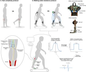
Example of how the smartphone app was used for this research. The top images show a black smartphone attached with a running arm band to the side or front of the shank - the two positions tested in this research. The middle figure shows the placement of the reflective markers for 3D motion analysis to evaluate the accuracy of the smartphone measurements. Markers were placed on the lateral epicondyle of the knee, lateral maleolus of the ankle, tibial tuberosity, and the distal tibia. Blacklight was used to mark the position of each marker and hide the position from the clinicians. The bottom panel shows screenshots from the app. The first screen is used to align the device and has arrows at the top and bottom that remind the clinician which anatomical landmarks should be used to align the device while displaying the shank-to-vertical angle in real time. The second screenshot shows an example of the calculated shank-to-vertical angle while someone was walking. The average is shown with a bold black line, with all other trials shown in blue and excluded trials (e.g., when someone was stopping or turning) that deviated more than one standard deviation from other trials are shown in red. There is also text below the graph that provides summary measures, like shank-to-vertical angle in mid stand and cadence (steps/min). The results can be exported as a picture or sent via e-mail using the app.
A) Smartphone positioning on the front or side of the shank. B) Reflective markers on the the tibial tuberosity (TT) – distal tibia (DT) and lateral epicondyle (LE) – lateral malleolus (LM) were used to compare the accuracy of the smartphone to traditional motion capture. UV markings were used to keep placement of these markers constant while blinding clinicians. C) Sample screenshots of the mobile application, including the set-up screen and results automatically produced after a walking trial.

Background

Assessments of human movement are clinically important. However, accurate measurements are often unavailable due to the need for expensive equipment or intensive processing. For orthotists and therapists, shank-to-vertical angle (SVA) is one critical measure used to assess gait and guide prescriptions. Smartphone-based sensors may provide a widely-available platform to expand access to quantitative assessments.

Objectives

Assess accuracy and repeatability of smartphone-based measurement of SVA compared to marker-based 3D motion analysis.

Method

Four licensed clinicians (two physical therapists and two orthotists) measured SVA during gait with a smartphone attached to the anterior or lateral shank surface of unimpaired adults.  We compared SVA calculated from the smartphone’s inertial measurement unit to marker-based measurements. Each clinician completed three sessions/day on two days with each participant to assess repeatability.

Results

Average absolute differences in SVA measured with a smartphone versus marker-based 3D motion analysis during gait were 0.67 ± 0.25° and 4.89 ± 0.72°, with anterior or lateral smartphone positions, respectively. The inter- and intra-day repeatability of SVA were within 2° for both smartphone positions.

Conclusions

Smartphone sensors can be used to measure SVA with high accuracy and repeatability during unimpaired gait, providing a widely-available tool for quantitative gait assessments.

Try it out!

The app for monitoring shank-to-vertical angle is available for you to download and use on either Android or iOS smartphone. Please complete THIS SURVEY which will then send you an e-mail with instructions for installation and use. This app is not an FDA approved medical device and should be used appropriately.

YC Pan, B Goodwin, E Sabelhaus, KM Peters, KF Bjornson, KLD Pham, WO Walker, KM Steele (2020) “Feasibility of using acceleration-derived jerk to quantify bimanual arm use” Journal of NeuroEngineering and Rehabilitation

Journal Article in Journal of NeuroEngineering & Rehabilitation

Two plots illustrating jerk ratio results. The plot on the left shows the probability distribution from one child with cerebral palsy before, during, and after constraint induced movement therapy. Before therapy, the probability distribution is shifted to the left of the center line, indicating that the individual relies much more on their non-paretic hand during daily life. During therapy, when their non-paretic hand is in a cast, the curve shifts to the right of the center line. This indicates they are using their paretic hand much more - which makes sense, since the other hand is in a cast. Unfortunately, after the cast is removed at the end of therapy, the curve is nearly identical to the curve before treatment, suggesting that after this intensive therapy the child did not use their paretic hand more during daily life. The figure on the right shows the summary metric from this plot, called jerk ratio 50 - which is just the 50% value of the probability density function - for all 5 children with cerebral palsy before, during, and after therapy. All the children have JR50 greater than 0.5 before therapy, which means they use their non-paretic hand more during daily life. During therapy, these values drop to 0.2 - 0.5, indicating that they use their paretic hand much more during CIMT. However, after therapy the JR50 values for all five participants return to close to their baseline value before therapy.
(Left) Example of jerk ratio distribution for one child with cerebral palsy before, during, and after constraint induced movement therapy. (Right) Summary metric of jerk ratio (jerk ratio-50) for all five children with cerebral palsy.

Background

Accelerometers have become common for evaluating the efficacy of rehabilitation for patients with neurologic disorders. For example, metrics like use ratio (UR) and magnitude ratio (MR) have been shown to differentiate movement patterns of children with cerebral palsy (CP) compared to typically-developing (TD) peers. However, these metrics are calculated from “activity counts” – a measure based on proprietary algorithms that approximate movement duration and intensity from raw accelerometer data. Algorithms used to calculate activity counts vary between devices, limiting comparisons of clinical and research results. The goal of this research was to develop complementary metrics based on raw accelerometer data to analyze arm movement after neurologic injury.

Method

We calculated jerk, the derivative of acceleration, to evaluate arm movement from accelerometer data. To complement current measures, we calculated jerk ratio (JR) as the relative jerk magnitude of the dominant (non-paretic) and non-dominant (paretic) arms.  We evaluated the JR distribution between arms and calculated the 50th percentile of the JR distribution (JR50). To evaluate these metrics, we analyzed bimanual accelerometry data for five children with hemiplegic CP who underwent Constraint-Induced Movement Therapy (CIMT) and five typically developing (TD) children. We compared JR between the CP and TD cohorts, and to activity count metrics.

Results

The JR50 differentiated between the CP and TD cohorts (CP = 0.578±0.041 before CIMT, TD = 0.506±0.026), demonstrating increased reliance on the non-dominant arm for the CP cohort. Jerk metrics also quantified changes in arm use during and after therapy (e.g., JR50 = 0.378±0.125 during CIMT, 0.591 ± 0.057 after CIMT). The JR was strongly correlated with UR and MR (r = -0.92, 0.89) for the CP cohort. For the TD cohort, JR50 was repeatable across three data collection periods with an average similarity of 0.945±0.015.

Conclusions

Acceleration-derived jerk captured differences in motion between TD and CP cohorts and correlated with activity count metrics. The code for calculating and plotting JR is open-source and available for others to use and build upon. By identifying device-independent metrics that can quantify arm movement in daily life, we hope to facilitate collaboration for rehabilitation research using wearable technologies.

Code

The algorithm for calculating jerk ratio, as well as user-friendly code to produce plots similar to the figure above are provided open-source as Python 3.6 code as a Python Jupyter Notebook within Google Colab. With this resource, research groups can use existing or newly created data from accelerometers to analyze jerk ratio as a complementary metric to existing measures, enabling comparison between research studies or centers that may rely on different sensors and activity count algorithms.

BR Shuman, M Goudriaan, K Desloovere, MH Schwartz, KM Steele (2019) “Muscle Synergy Constraints Do Not Improve Estimates of Muscle Activity From Static Optimization During Gait for Unimpaired Children or Children With Cerebral Palsy” Frontiers in Neurorobotics

Journal Article in Frontiers of Neurorobotics:

This study demonstrated that muscle activations estimated from static optimization using generic musculoskeletal modeling does not accurately predict EMG profiles for children with CP or TD peers. Constraining activation patterns to experimentally measured synergies increased estimated muscle stresses, but did not improve the estimation of muscle activations for either group.

figure depicting flow chart with modeling optimization and muscle activity
Constraining simulated activations in inverse dynamic simulations to subject-specific synergies alone does not improve estimation of muscle activations during gait for generic musculoskeletal models.

Background

Neuromusculoskeletal simulation provides a promising platform to inform the design of assistive devices or inform rehabilitation. For these applications, a simulation must be able to accurately represent the person of interest, such as an individual with a neurologic injury. If a simulation fails to predict how an individual recruits and coordinates their muscles during movement, it will have limited utility for informing design or rehabilitation. While inverse dynamic simulations have previously been used to evaluate anticipated responses from interventions, like orthopaedic surgery or orthoses, they frequently struggle to accurately estimate muscle activations, even for tasks like walking. The simulated muscle activity often fails to represent experimentally measured muscle activity from electromyographic (EMG) recordings. Research has theorized that the nervous system may simplify the range of possible activations used during dynamic tasks, by constraining activations to weighted groups of muscles, referred to as muscle synergies. Synergies are altered after neurological injury, such as stroke or cerebral palsy (CP), and may provide a method for improving subject-specific models of neuromuscular control.

Purpose

The aim of this study was to test whether constraining simulation to synergies could improve estimated muscle activations compared to EMG data.

Method

We evaluated modeled muscle activations during gait for six typically developing children (TD) and six children with CP. Muscle activations were estimated with: 1) static optimization (SO), minimizing muscle activations squared, and 2) synergy static optimization (SynSO), minimizing synergy activations squared using the weights identified from EMG data for 2-5 synergies.

Results

While SynSO caused changes in estimated activations compared to SO, the correlation to EMG data was not higher in SynSO than SO for either TD or CP groups . The correlations to EMG were higher in CP than TD for both SO (CP: 0.48, TD: 0.36) and SynSO (CP: 0.46, TD: 0.26 for 5 synergies). Constraining activations to SynSO caused the simulated muscle stress to increase compared to SO for all individuals, causing a 157% increase with two synergies.

Conclusions

These results suggest that constraining simulated activations in inverse dynamic simulations to subject-specific synergies alone does not improve estimation of muscle activations during gait for generic musculoskeletal models.

ESMAC 2019: Award Finalists

Congratulations to Nicole Zaino and our colleague Mike Schwartz at Gillette Children’s Specialty Healthcare for both being nominated as finalists for the Best Presentation Award at the upcoming ESMAC Conference in Amsterdam. Their abstracts are among the top 16 submissions to the conference and the final award will be determined based upon their presentations.

Logo for the 2019 ESMAC meeting overlaid on a classic Amsterdam scene, bikes lined up on a bridge over a canal with historic buildings in the background.

Nicole will be presenting her research:

Spasticity reduction in children with cerebral palsy is not associated with reduced energy during walking 

Selective dorsal rhizotomy reduces spasticity, but does it also reduce energy consumption during walking? In an analysis of over 300 children with cerebral palsy, Nicole demonstrated that although rhiztomy does reduce spasticity, it does not reduce energy consumption. These results provide further evidence that spasticity is not a main contributor to elevated energy among people with cerebral palsy. You can also learn more about this study from our recent submitted manuscript, available on bioRxiv.

Mike will be presenting his research:

The effects of walking speed and age on energy consumption in children with cerebral palsy and their typically developing peers

We know that walking energy is high among people with cerebral palsy, and that energy varies with speed and age. Using retrospective data of over 300 kids with cerebral palsy and 150 typically-developing peers, Mike used a statistical model to evaluate these speed and age effects. He found that energy decreases until 8-10 years of age for kids with CP, while it remains stable beyond age 5 for typically-developing peers. Kids with CP also have a greater elevation in energy with greater walking speeds. These results are important to help quantify and understand impacts of interventions, like surgery or assistive devices, which are often done during this time period when kids are still growing and developing.

They will both be presenting in the Optimizing Energy Cost session from 11:40-12:30 on Thursday, September 26th.

Best of luck to Nicole & Mike!

Research Experience Undergraduates Present at CNT

This summer the Steele Lab had the pleasure of hosting three undergraduate researchers – Robin Yan from University of Washington, Ava Lakmazaheri from Olin College of Engineering, and Katherine Chamblin from University of Washington.

After a competitive selection process, students are offered a 10-week internship here at the University to work directly with a research lab on campus. One of the program’s final deliverables is a presentation of their work, both in podium and poster format, to members of the local and scientific community. Congratulations to Robin, Ava, and Katherine for their successful time here in the lab, and for giving polished presentations.

Group of six individuals standing shoulder to shoulder and smiling in front of white wall
REU Students with their lab mentors

Robin examined biomechanical analyses of typically developing individuals during emulation of cerebral palsy gait and Ava worked on optimizing musculoskeletal models for children with cerebral palsy.

Sun shinning down on young woman in business attire talking to another woman in front of a white and purple poster board
Katherine discussing her work with an interested student

Katherine investigated social communication patterns of children with cerebral palsy and their families after integrating an early-powered mobility device