Join us for the following AccessEngineering Webinars! If you would like to attend one or more, please register for the respective topic by following the link provided. (Please note, all of the webinars are scheduled in Eastern Daylight Time)
Choosing and Delivering a High Quality Online Program
Friday, Dec 4th, 2015 11:00 am – 12:00 pm (EDT)
About the Webinar:
This webinar will focus on three aspects of distance education: (1) how Kettering University Online develops high quality online programs; (2) how can students choose a high quality online program to enroll; and (3) how can potential instructors find a high quality institution to teach. There will be presentations and Q&A sessions about each aspect.
Presenter: Dr. Christine Wallace
AccessEngineering: Strategies to support individuals with disabilities pursuing careers in engineering – Part 1
Monday, December 7th, 2015 1:00 pm – 2:00 pm (EDT)
About the Webinar:
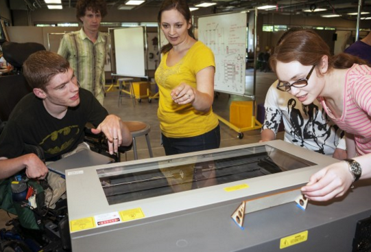
This webinar focuses on a new NSF initiative, AccessEngineering, which (1) supports and promotes individuals with disabilities in pursuing engineering, and (2) integrates universal design and accessibility topics into the engineering curriculum. In particular, this webinar will discuss topics including communication tips and how you can best support individuals with disabilities in engineering, and best practices for making makerspaces, labs and machine shops accessible.
Presenters: Sheryl Burgstahler, Richard Ladner, Maya Cakmak, Kat Steele, Brianna Blaser
Patents: An Introduction for Inventors, Part One
Friday, December 11th, 2015 2:00 pm – 3:00 pm (EDT)
About the Webinar:
This presentation series is a basic introduction to patents and an overview of issues and practices to be considered by prospective patent inventors and patent owners. Part One focuses on providing a basic overview of patents and need-to-know information for inventors to streamline the patenting process. The basic overview explains what patents are, how patents relate to other forms of intellectual property, what requirements must be met for patenting an invention in the U.S., basic considerations for patenting an invention internationally, and how patents provide value to their owners. The overview of information for inventors covers inventorship and ownership issues, when to file for a patent, how to avoid sinking a patent application with one’s own publications, and various good practices for inventors to follow regarding disclosures and record-keeping.
Presenter: Michael Gamble
AccessEngineering: Strategies to support individuals with disabilities pursuing careers in engineering – Part 2
Tuesday, January 19th, 2016 1:00 pm – 2:00 pm (EDT)
About the Webinar:
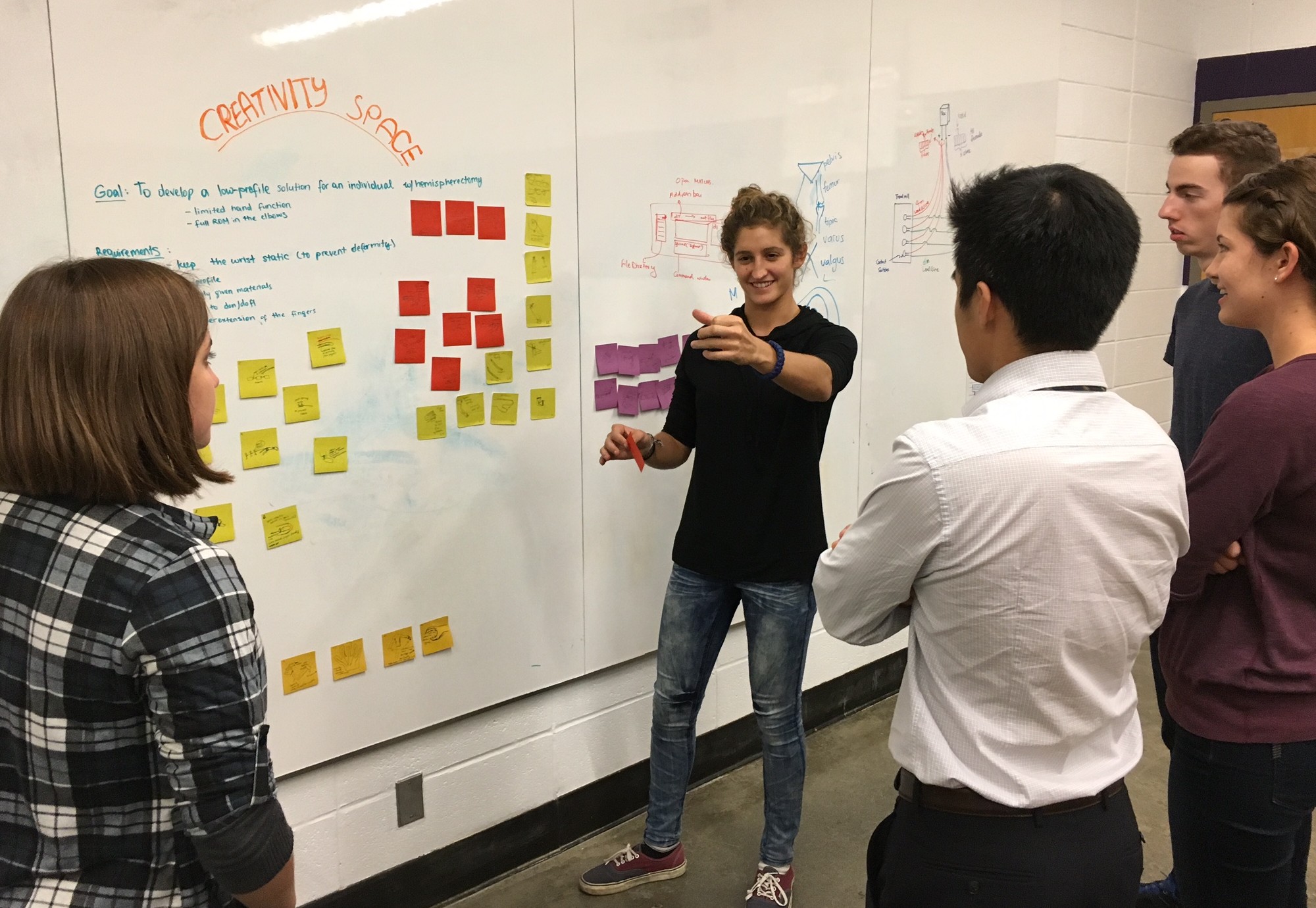
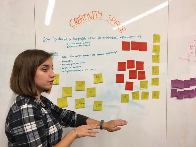
This webinar focuses on a new NSF initiative, AccessEngineering, which (1) supports and promotes individuals with disabilities in pursuing engineering, and (2) integrates universal design and accessibility topics into the engineering curriculum. In particular, this webinar will discuss topics including communication tips and how you can best support individuals with disabilities in engineering, and best practices for making makerspaces, labs and machine shops accessible. Part 2 of this webinar series will focus on strategies for easily integrating universal design and accessibility topics into engineering education.
Presenters: Sheryl Burgstahler, Richard Ladner, Maya Cakmak, Kat Steele
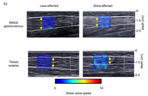
 “Use of shear wave ultrasound elastography to quantify muscle properties in cerebral palsy.” Clinical Biomechanics")